Detailed DSSR results for the G-quadruplex: PDB entry 6z8w
Created and maintained by Xiang-Jun Lu <xiangjun@x3dna.org>
Citation: Please cite the NAR'20 DSSR-PyMOL schematics paper and/or the NAR'15 DSSR method paper.
Summary information
- PDB id
- 6z8w
- Class
- hydrolase
- Method
- X-ray (1.73 Å)
- Summary
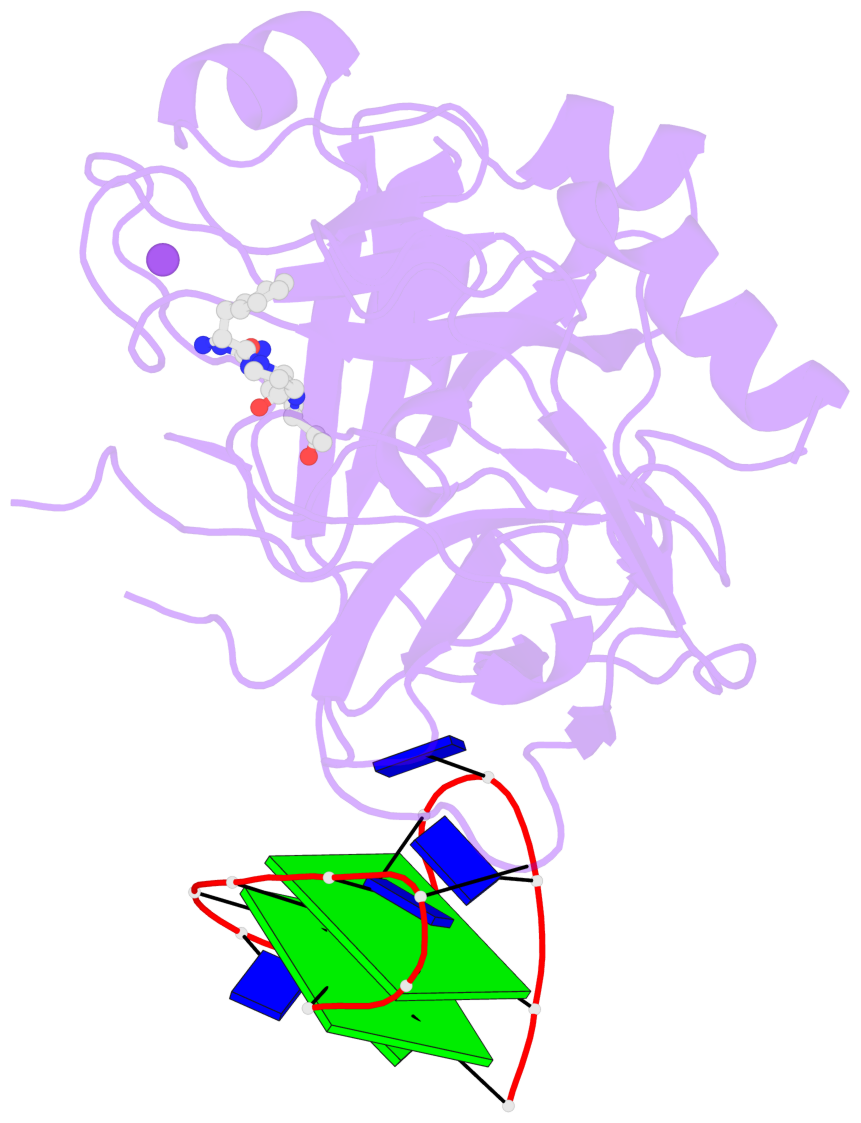
- X-ray structure of the complex between human alpha thrombin and a thrombin binding aptamer variant (tba-3g), which contains 1-beta-d-glucopyranosyl residue in the side chain of thy3 at n3.
- Reference
- Smirnov I, Kolganova N, Troisi R, Sica F, Timofeev E (2021): "Expanding the recognition interface of the thrombin-binding aptamer HD1 through modification of residues T3 and T12." Mol Ther Nucleic Acids, 23, 863-871. doi: 10.1016/j.omtn.2021.01.004.
- Abstract
- Post-SELEX modification of DNA aptamers is an established strategy to improve their affinity or inhibitory characteristics. In this study, we examined the possibility of increasing the recognition interface between the thrombin-binding aptamer HD1 (TBA) and thrombin by adding a chemically modified side chain to selected nucleotide residues. A panel of 22 TBA variants with N3-modified residues T3 and T12 was prepared by a two-step modification procedure. Aptamers were characterized by a combination of biophysical and biochemical methods. We identified mutants with enhanced affinity and improved anticoagulant activity. The crystal structures of thrombin complexes with three selected modified variants revealed that the modified pyrimidine base invariably allocates in proximity to thrombin residues Tyr76 and Ile82 due to the directing role of the unmodified TT loop. The modifications induced an increase in the contact areas between thrombin and the modified TBAs. Comparative analysis of the structural, biochemical, and biophysical data suggests that the non-equivalent binding modes of the mutants with thrombin in the T3- and T12-modified series account for the observed systematic differences in their affinity characteristics. In this study, we show that extending the recognition surface between the protein and modified aptamers is a promising approach that may improve characteristics of aptamer ligands.
- G4 notes
- 2 G-tetrads, 1 G4 helix, 1 G4 stem, 2(+Ln+Lw+Ln), chair(2+2), UDUD
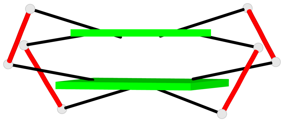
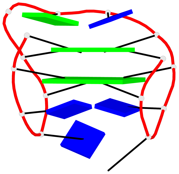
Base-block schematics in six views
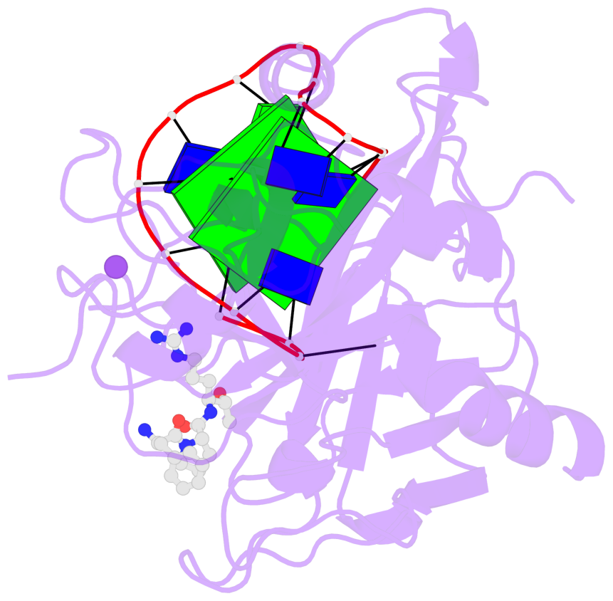
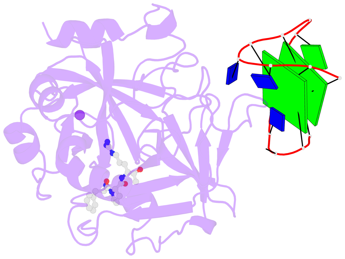

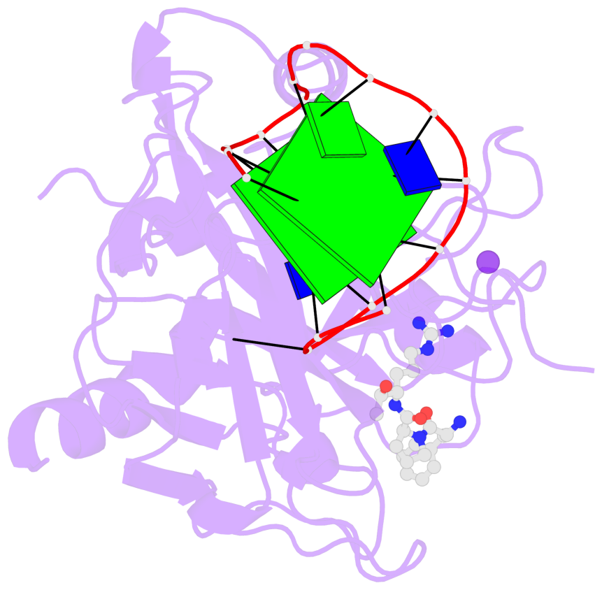
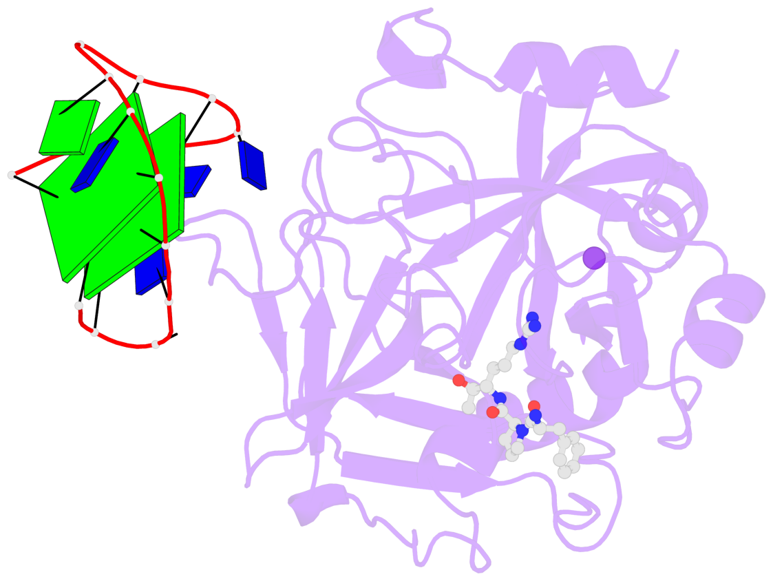
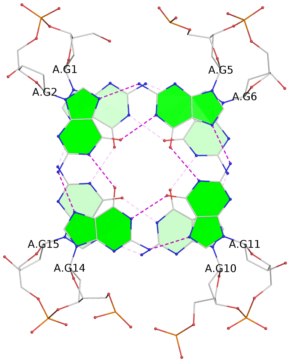
List of 2 G-tetrads
 1 glyco-bond=s-s- sugar=---- groove=wnwn planarity=0.519 type=saddle nts=4 GGGG A.DG1,A.DG15,A.DG10,A.DG6
 2 glyco-bond=-s-s sugar=---- groove=wnwn planarity=0.572 type=other  nts=4 GGGG A.DG2,A.DG14,A.DG11,A.DG5
List of 1 G4-helix
In DSSR, a G4-helix is defined by stacking interactions of G-tetrads, regardless of backbone connectivity, and may contain more than one G4-stem.
Helix#1, 2 G-tetrad layers, INTRA-molecular, with 1 stem
 |
1 glyco-bond=s-s- sugar=---- groove=wnwn Major-->WC nts=4 GGGG A.DG1,A.DG15,A.DG10,A.DG6 2 glyco-bond=-s-s sugar=---- groove=wnwn WC-->Major nts=4 GGGG A.DG2,A.DG14,A.DG11,A.DG5 step#1 mm(<>,outward) area=15.96 rise=3.51 twist=15.8 strand#1 DNA glyco-bond=s- sugar=-- nts=2 GG A.DG1,A.DG2 strand#2 DNA glyco-bond=-s sugar=-- nts=2 GG A.DG15,A.DG14 strand#3 DNA glyco-bond=s- sugar=-- nts=2 GG A.DG10,A.DG11 strand#4 DNA glyco-bond=-s sugar=-- nts=2 GG A.DG6,A.DG5 |
| 1 stacking diagram | |
 |
1 glyco-bond=s-s- sugar=---- groove=wnwn Major-->WC nts=4 GGGG A.DG1,A.DG15,A.DG10,A.DG6 |
List of 1 G4-stem
In DSSR, a G4-stem is defined as a G4-helix with backbone connectivity. Bulges are also allowed along each of the four strands.
Stem#1, 2 G-tetrad layers, 3 loops, INTRA-molecular, UDUD, anti-parallel, 2(+Ln+Lw+Ln), chair(2+2)
 |
1 glyco-bond=s-s- sugar=---- groove=wnwn Major-->WC nts=4 GGGG A.DG1,A.DG15,A.DG10,A.DG6 2 glyco-bond=-s-s sugar=---- groove=wnwn WC-->Major nts=4 GGGG A.DG2,A.DG14,A.DG11,A.DG5 step#1 mm(<>,outward) area=15.96 rise=3.51 twist=15.8 strand#1 U DNA glyco-bond=s- sugar=-- nts=2 GG A.DG1,A.DG2 strand#2 D DNA glyco-bond=-s sugar=-- nts=2 GG A.DG15,A.DG14 strand#3 U DNA glyco-bond=s- sugar=-- nts=2 GG A.DG10,A.DG11 strand#4 D DNA glyco-bond=-s sugar=-- nts=2 GG A.DG6,A.DG5 loop#1 type=lateral strands=[#1,#4] nts=2 aT A.QCK3,A.DT4 loop#2 type=lateral strands=[#4,#3] nts=3 TGT A.DT7,A.DG8,A.DT9 loop#3 type=lateral strands=[#3,#2] nts=2 TT A.DT12,A.DT13 |