Detailed DSSR results for the G-quadruplex: PDB entry 7d31
Created and maintained by Xiang-Jun Lu <xiangjun@x3dna.org>
Citation: Please cite the NAR'20 DSSR-PyMOL schematics paper and/or the NAR'15 DSSR method paper.
Summary information
- PDB id
- 7d31
- Class
- DNA
- Method
- X-ray (1.396 Å)
- Summary
- The tba-pb2+ complex in p41212 space group
- Reference
- Liu H, Gao Y, Mathivanan J, Shen F, Chen X, Li Y, Shao Z, Zhang Y, Shao Q, Sheng J, Gan J (2022): "Structure-guided development of Pb 2+ -binding DNA aptamers." Sci Rep, 12, 460. doi: 10.1038/s41598-021-04243-2.
- Abstract
- Owing to its great threat to human health and environment, Pb2+ pollution has been recognized as a major public problem by the World Health Organization (WHO). Many DNA aptamers have been utilized in the development of Pb2+-detection sensors, but the underlying mechanisms remain elusive. Here, we report three Pb2+-complexed structures of the thrombin binding aptamer (TBA). These high-resolution crystal structures showed that TBA forms intramolecular G-quadruplex and Pb2+ is bound by the two G-tetrads in the center. Compared to K+-stabilized G-quadruplexes, the coordinating distance between Pb2+ and the G-tetrads are much shorter. The T3T4 and T12T13 linkers play important roles in dimerization and crystallization of TBA, but they are changeable for Pb2+-binding. In combination with mutagenesis and CD spectra, the G8C mutant structure unraveled that the T7G8T9 linker of TBA is also variable. In addition to expansion of the Pb2+-binding aptamer sequences, our study also set up one great example for quick and rational development of other aptamers with similar or optimized binding activity.
- G4 notes
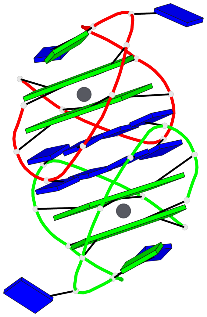
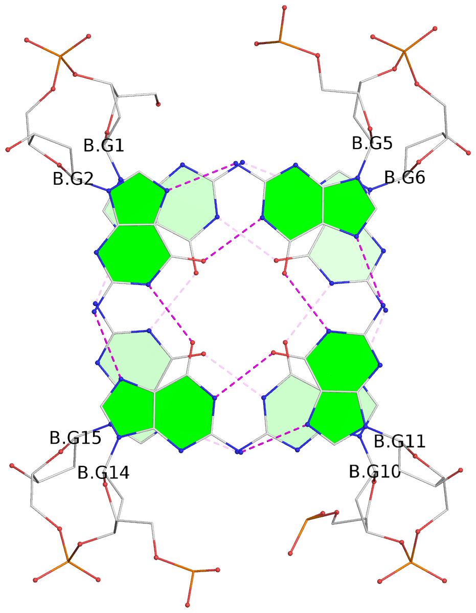
- 4 G-tetrads, 2 G4 helices, 2 G4 stems, 2(+Ln+Lw+Ln), chair(2+2), UDUD
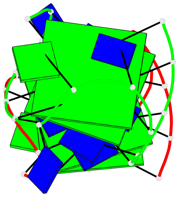
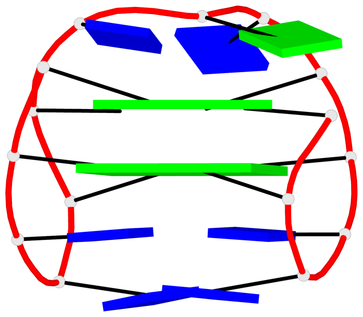
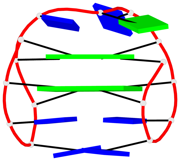
Base-block schematics in six views
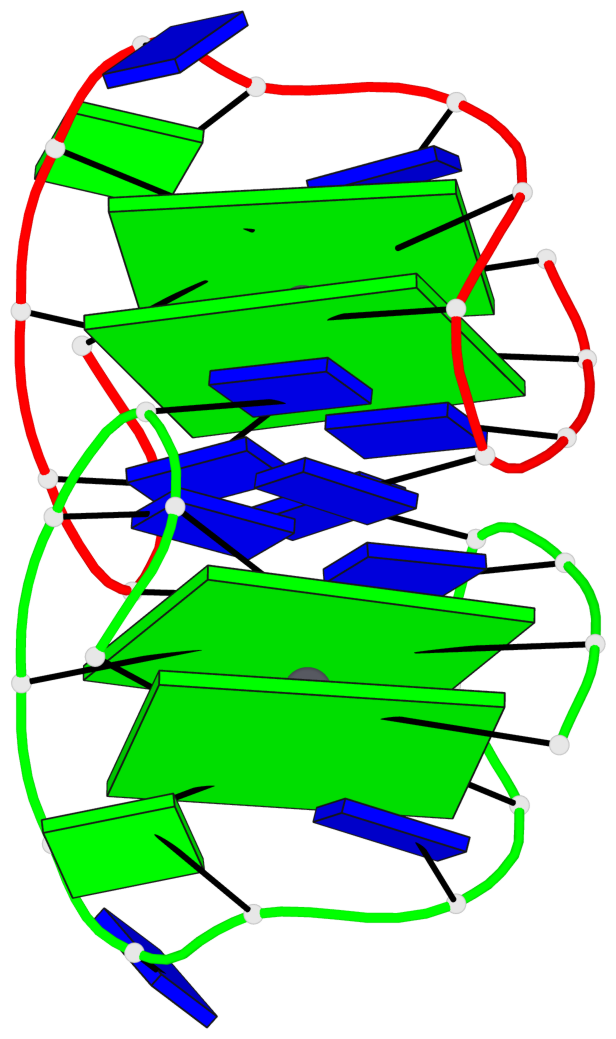
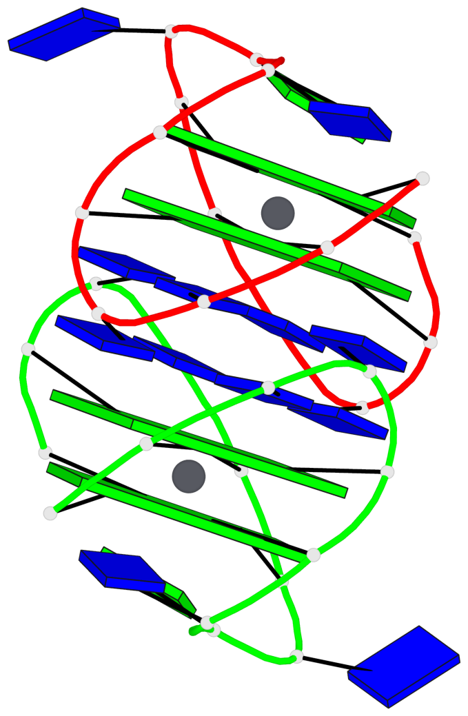
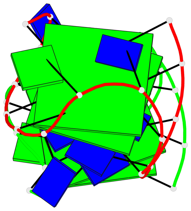
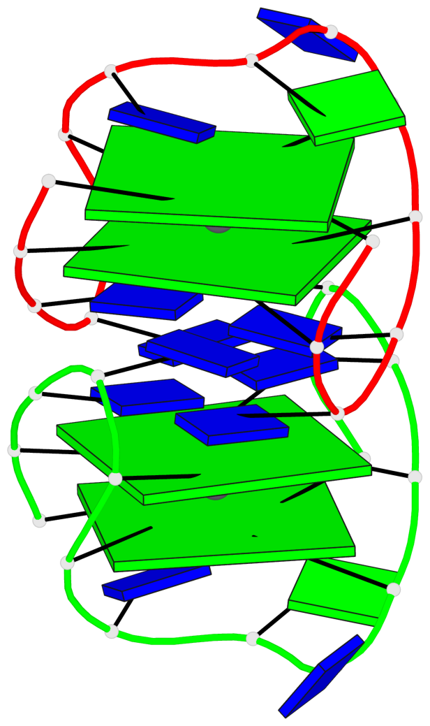
List of 4 G-tetrads
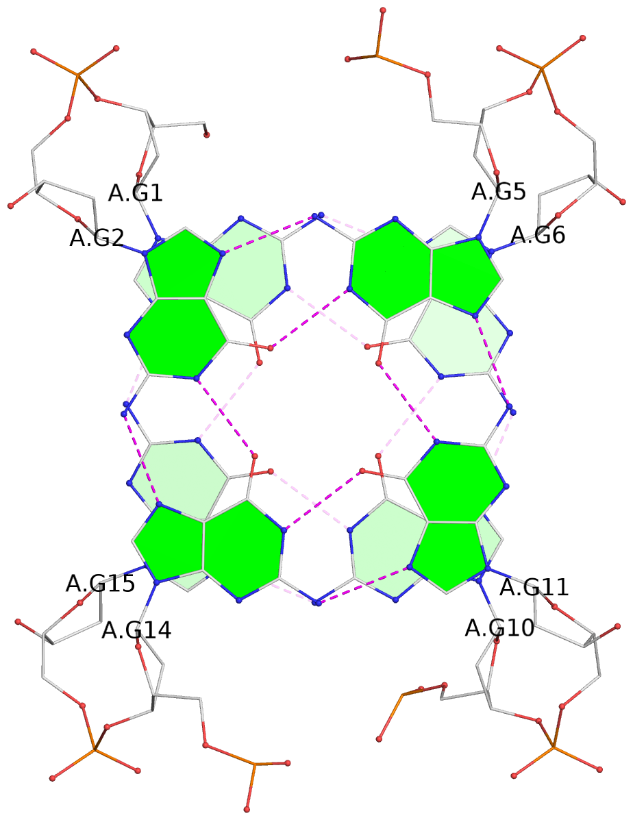
 1 glyco-bond=s-s- sugar=---- groove=wnwn planarity=0.451 type=bowl-2 nts=4 GGGG A.DG1,A.DG15,A.DG10,A.DG6
 2 glyco-bond=-s-s sugar=---- groove=wnwn planarity=0.417 type=other  nts=4 GGGG A.DG2,A.DG14,A.DG11,A.DG5
 3 glyco-bond=s-s- sugar=---- groove=wnwn planarity=0.443 type=bowl-2 nts=4 GGGG B.DG1,B.DG15,B.DG10,B.DG6
 4 glyco-bond=-s-s sugar=---- groove=wnwn planarity=0.422 type=bowl-2 nts=4 GGGG B.DG2,B.DG14,B.DG11,B.DG5
List of 2 G4-helices
In DSSR, a G4-helix is defined by stacking interactions of G-tetrads, regardless of backbone connectivity, and may contain more than one G4-stem.
Helix#1, 2 G-tetrad layers, INTRA-molecular, with 1 stem
 |
1 glyco-bond=s-s- sugar=---- groove=wnwn Major-->WC nts=4 GGGG A.DG1,A.DG15,A.DG10,A.DG6 2 glyco-bond=-s-s sugar=---- groove=wnwn WC-->Major nts=4 GGGG A.DG2,A.DG14,A.DG11,A.DG5 step#1 mm(<>,outward) area=18.29 rise=3.49 twist=11.8 strand#1 DNA glyco-bond=s- sugar=-- nts=2 GG A.DG1,A.DG2 strand#2 DNA glyco-bond=-s sugar=-- nts=2 GG A.DG15,A.DG14 strand#3 DNA glyco-bond=s- sugar=-- nts=2 GG A.DG10,A.DG11 strand#4 DNA glyco-bond=-s sugar=-- nts=2 GG A.DG6,A.DG5 |
| 1 stacking diagram | |
 |
1 glyco-bond=s-s- sugar=---- groove=wnwn Major-->WC nts=4 GGGG A.DG1,A.DG15,A.DG10,A.DG6 |
Helix#2, 2 G-tetrad layers, INTRA-molecular, with 1 stem
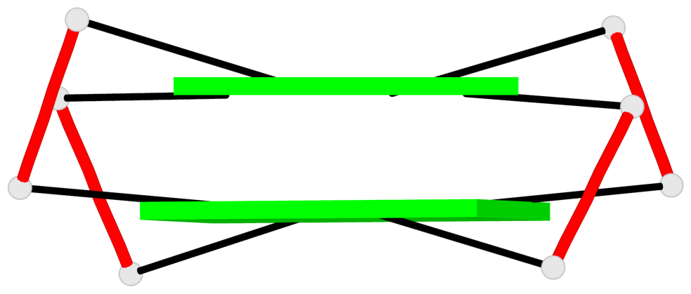 |
1 glyco-bond=s-s- sugar=---- groove=wnwn Major-->WC nts=4 GGGG B.DG1,B.DG15,B.DG10,B.DG6 2 glyco-bond=-s-s sugar=---- groove=wnwn WC-->Major nts=4 GGGG B.DG2,B.DG14,B.DG11,B.DG5 step#1 mm(<>,outward) area=17.84 rise=3.49 twist=12.3 strand#1 DNA glyco-bond=s- sugar=-- nts=2 GG B.DG1,B.DG2 strand#2 DNA glyco-bond=-s sugar=-- nts=2 GG B.DG15,B.DG14 strand#3 DNA glyco-bond=s- sugar=-- nts=2 GG B.DG10,B.DG11 strand#4 DNA glyco-bond=-s sugar=-- nts=2 GG B.DG6,B.DG5 |
| 1 stacking diagram | |
 |
1 glyco-bond=s-s- sugar=---- groove=wnwn Major-->WC nts=4 GGGG B.DG1,B.DG15,B.DG10,B.DG6 |
List of 2 G4-stems
In DSSR, a G4-stem is defined as a G4-helix with backbone connectivity. Bulges are also allowed along each of the four strands.
Stem#1, 2 G-tetrad layers, 3 loops, INTRA-molecular, UDUD, anti-parallel, 2(+Ln+Lw+Ln), chair(2+2)
 |
1 glyco-bond=s-s- sugar=---- groove=wnwn Major-->WC nts=4 GGGG A.DG1,A.DG15,A.DG10,A.DG6 2 glyco-bond=-s-s sugar=---- groove=wnwn WC-->Major nts=4 GGGG A.DG2,A.DG14,A.DG11,A.DG5 step#1 mm(<>,outward) area=18.29 rise=3.49 twist=11.8 strand#1 U DNA glyco-bond=s- sugar=-- nts=2 GG A.DG1,A.DG2 strand#2 D DNA glyco-bond=-s sugar=-- nts=2 GG A.DG15,A.DG14 strand#3 U DNA glyco-bond=s- sugar=-- nts=2 GG A.DG10,A.DG11 strand#4 D DNA glyco-bond=-s sugar=-- nts=2 GG A.DG6,A.DG5 loop#1 type=lateral strands=[#1,#4] nts=2 TT A.DT3,A.DT4 loop#2 type=lateral strands=[#4,#3] nts=3 TGT A.DT7,A.DG8,A.DT9 loop#3 type=lateral strands=[#3,#2] nts=2 TT A.DT12,A.DT13 |
Stem#2, 2 G-tetrad layers, 3 loops, INTRA-molecular, UDUD, anti-parallel, 2(+Ln+Lw+Ln), chair(2+2)
 |
1 glyco-bond=s-s- sugar=---- groove=wnwn Major-->WC nts=4 GGGG B.DG1,B.DG15,B.DG10,B.DG6 2 glyco-bond=-s-s sugar=---- groove=wnwn WC-->Major nts=4 GGGG B.DG2,B.DG14,B.DG11,B.DG5 step#1 mm(<>,outward) area=17.84 rise=3.49 twist=12.3 strand#1 U DNA glyco-bond=s- sugar=-- nts=2 GG B.DG1,B.DG2 strand#2 D DNA glyco-bond=-s sugar=-- nts=2 GG B.DG15,B.DG14 strand#3 U DNA glyco-bond=s- sugar=-- nts=2 GG B.DG10,B.DG11 strand#4 D DNA glyco-bond=-s sugar=-- nts=2 GG B.DG6,B.DG5 loop#1 type=lateral strands=[#1,#4] nts=2 TT B.DT3,B.DT4 loop#2 type=lateral strands=[#4,#3] nts=3 TGT B.DT7,B.DG8,B.DT9 loop#3 type=lateral strands=[#3,#2] nts=2 TT B.DT12,B.DT13 |