Detailed DSSR results for the G-quadruplex: PDB entry 7zkn
Created and maintained by Xiang-Jun Lu <xiangjun@x3dna.org>
Citation: Please cite the NAR'20 DSSR-PyMOL schematics paper and/or the NAR'15 DSSR method paper.
Summary information
- PDB id
- 7zkn
- Class
- hydrolase
- Method
- X-ray (3.03 Å)
- Summary
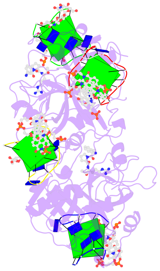
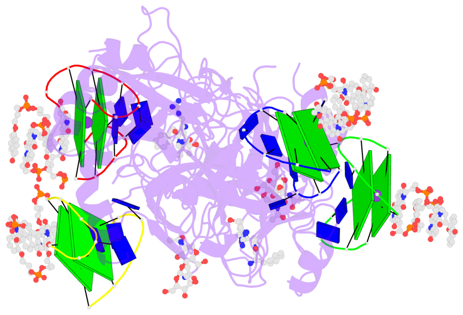
- X-ray structure of the complex between human alpha thrombin and a pseudo-cyclic thrombin binding aptamer (tba-nnp-ddp) - crystal form gamma
- Reference
- Troisi R, Riccardi C, Perez de Carvasal K, Smietana M, Morvan F, Del Vecchio P, Montesarchio D, Sica F (2022): "A terminal functionalization strategy reveals unusual binding abilities of anti-thrombin anticoagulant aptamers." Mol Ther Nucleic Acids, 30, 585-594. doi: 10.1016/j.omtn.2022.11.007.
- Abstract
- Despite their unquestionable properties, oligonucleotide aptamers display some drawbacks that continue to hinder their applications. Several strategies have been undertaken to overcome these weaknesses, using thrombin binding aptamers as proof-of-concept. In particular, the functionalization of a thrombin exosite I binding aptamer (TBA) with aromatic moieties, e.g., naphthalene dimides (N) and dialkoxynaphthalenes (D), attached at the 5' and 3' ends, respectively, proved to be highly promising. To obtain a molecular view of the effects of these modifications on aptamers, we performed a crystallographic analysis of one of these engineered oligonucleotides (TBA-NNp/DDp) in complex with thrombin. Surprisingly, three of the four examined crystallographic structures are ternary complexes in which thrombin binds a TBA-NNp/DDp molecule at exosite II as well as at exosite I, highlighting the ability of this aptamer, differently from unmodified TBA, to also recognize a localized region of exosite II. This novel ability is strictly related to the solvophobic behavior of the terminal modifications. Studies were also performed in solution to examine the properties of TBA-NNp/DDp in a crystal-free environment. The present results throw new light on the importance of appendages inducing a pseudo-cyclic charge-transfer structure in nucleic acid-based ligands to improve the interactions with proteins, thus considerably widening their potentialities.
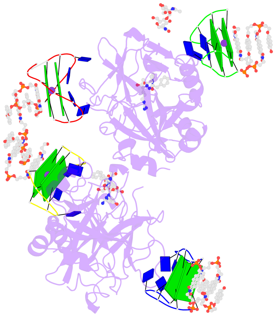
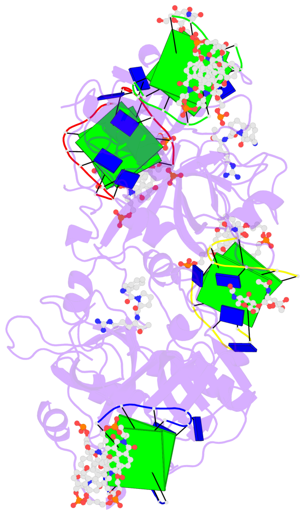
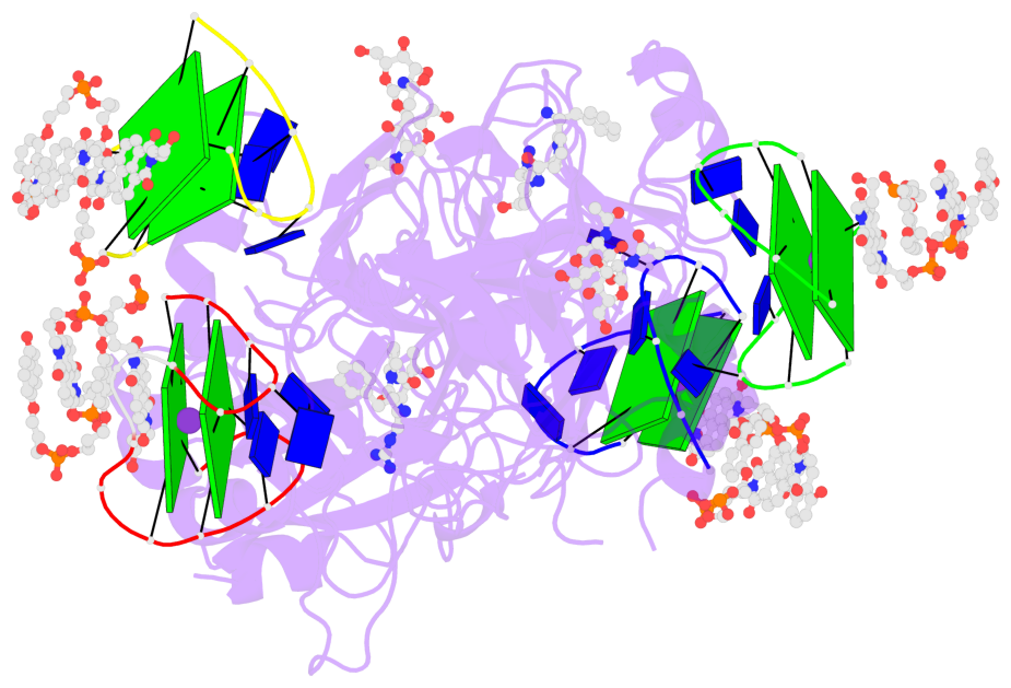
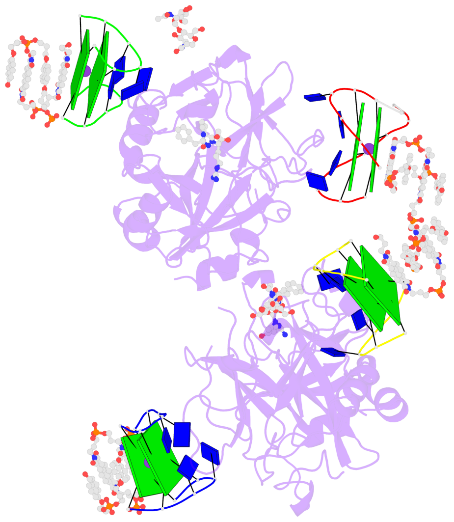
- G4 notes
- 8 G-tetrads, 4 G4 helices, 4 G4 stems, 2(+Ln+Ln), (2+2), UDUD; 2(+Ln+Lw+Ln), chair(2+2), UDUD
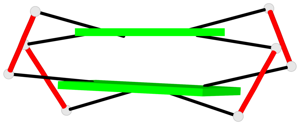
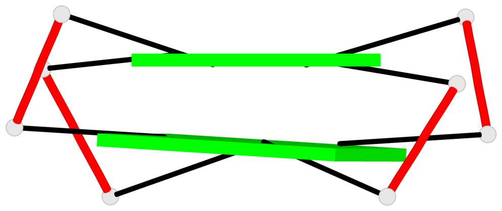
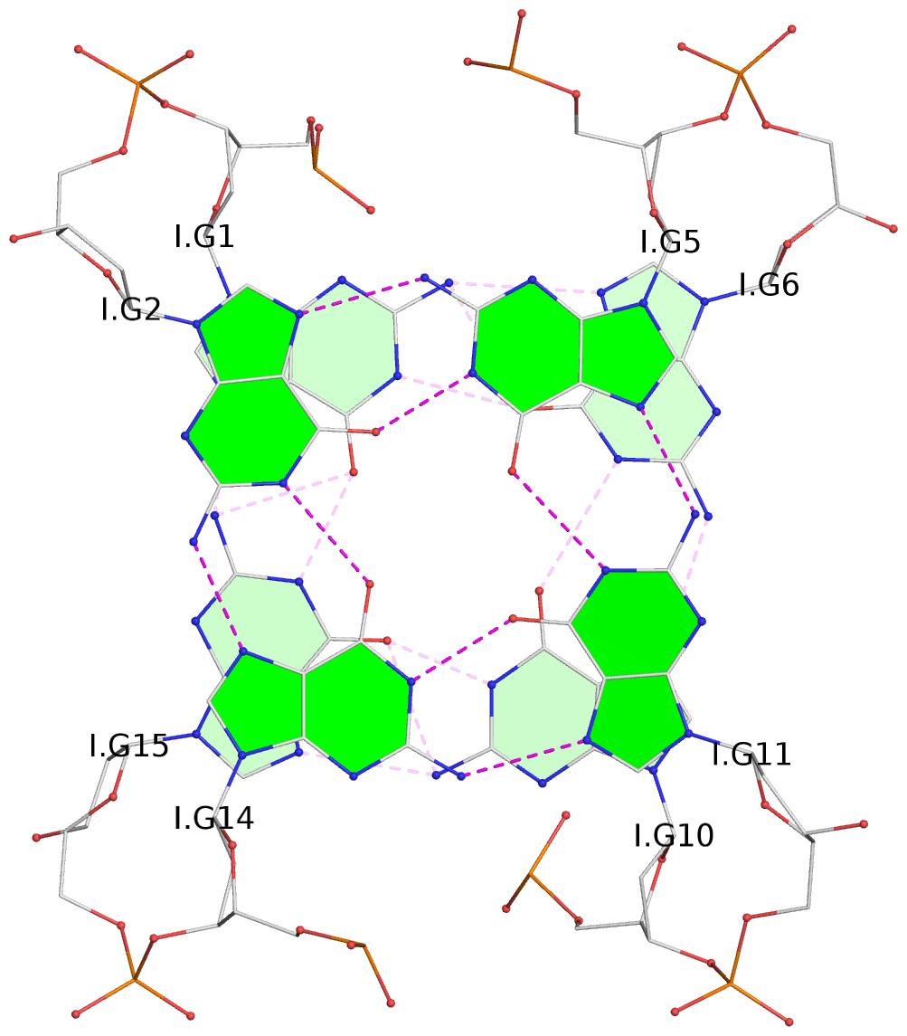
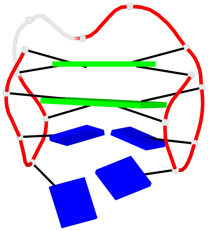
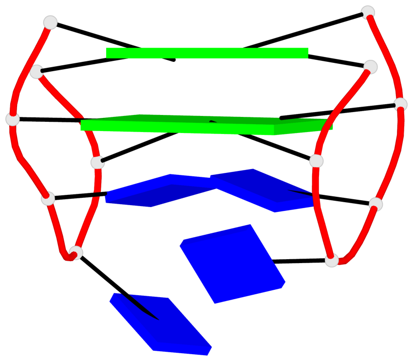
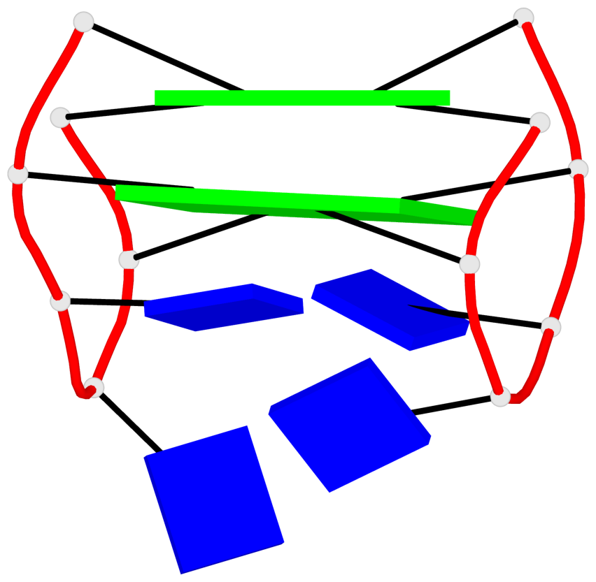
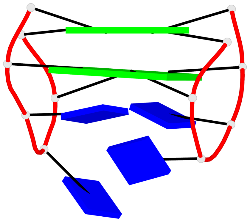
Base-block schematics in six views
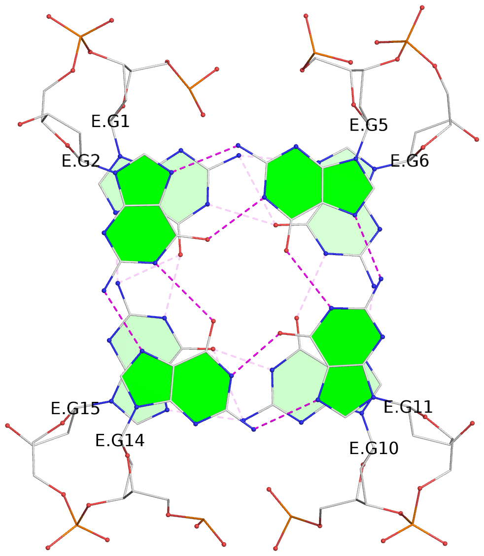
List of 8 G-tetrads
 1 glyco-bond=s-s- sugar=---- groove=wnwn planarity=0.350 type=other  nts=4 GGGG E.DG1,E.DG15,E.DG10,E.DG6
 2 glyco-bond=-s-s sugar=---- groove=wnwn planarity=0.415 type=bowl-2 nts=4 GGGG E.DG2,E.DG14,E.DG11,E.DG5
 3 glyco-bond=s-s- sugar=-3-. groove=wnwn planarity=0.175 type=other  nts=4 GGGG F.DG1,F.DG15,F.DG10,F.DG6
 4 glyco-bond=-s-s sugar=--.- groove=wnwn planarity=0.308 type=bowl-2 nts=4 GGGG F.DG2,F.DG14,F.DG11,F.DG5
 5 glyco-bond=s-s- sugar=-3-. groove=wnwn planarity=0.477 type=other  nts=4 GGGG G.DG1,G.DG15,G.DG10,G.DG6
 6 glyco-bond=-s-s sugar=.--- groove=wnwn planarity=0.463 type=bowl-2 nts=4 GGGG G.DG2,G.DG14,G.DG11,G.DG5
 7 glyco-bond=s-s- sugar=-3-- groove=wnwn planarity=0.315 type=saddle nts=4 GGGG I.DG1,I.DG15,I.DG10,I.DG6
 8 glyco-bond=-s-s sugar=.-.- groove=wnwn planarity=0.357 type=other  nts=4 GGGG I.DG2,I.DG14,I.DG11,I.DG5
List of 4 G4-helices
In DSSR, a G4-helix is defined by stacking interactions of G-tetrads, regardless of backbone connectivity, and may contain more than one G4-stem.
Helix#1, 2 G-tetrad layers, INTRA-molecular, with 1 stem
 |
1 glyco-bond=s-s- sugar=---- groove=wnwn Major-->WC nts=4 GGGG E.DG1,E.DG15,E.DG10,E.DG6 2 glyco-bond=-s-s sugar=---- groove=wnwn WC-->Major nts=4 GGGG E.DG2,E.DG14,E.DG11,E.DG5 step#1 mm(<>,outward) area=19.44 rise=3.59 twist=14.7 strand#1 DNA glyco-bond=s- sugar=-- nts=2 GG E.DG1,E.DG2 strand#2 DNA glyco-bond=-s sugar=-- nts=2 GG E.DG15,E.DG14 strand#3 DNA glyco-bond=s- sugar=-- nts=2 GG E.DG10,E.DG11 strand#4 DNA glyco-bond=-s sugar=-- nts=2 GG E.DG6,E.DG5 |
| 1 stacking diagram | |
 |
1 glyco-bond=s-s- sugar=---- groove=wnwn Major-->WC nts=4 GGGG E.DG1,E.DG15,E.DG10,E.DG6 |
Helix#2, 2 G-tetrad layers, INTRA-molecular, with 1 stem
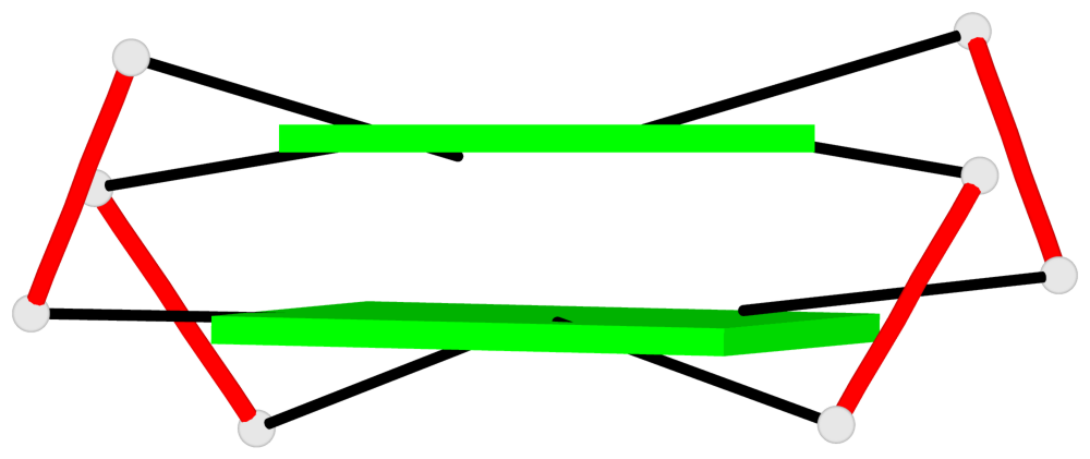 |
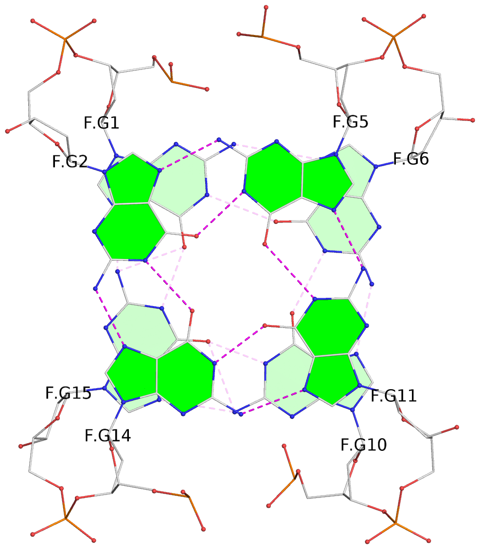
1 glyco-bond=s-s- sugar=-3-. groove=wnwn Major-->WC nts=4 GGGG F.DG1,F.DG15,F.DG10,F.DG6 2 glyco-bond=-s-s sugar=--.- groove=wnwn WC-->Major nts=4 GGGG F.DG2,F.DG14,F.DG11,F.DG5 step#1 mm(<>,outward) area=16.82 rise=3.40 twist=16.9 strand#1 DNA glyco-bond=s- sugar=-- nts=2 GG F.DG1,F.DG2 strand#2 DNA glyco-bond=-s sugar=3- nts=2 GG F.DG15,F.DG14 strand#3 DNA glyco-bond=s- sugar=-. nts=2 GG F.DG10,F.DG11 strand#4 DNA glyco-bond=-s sugar=.- nts=2 GG F.DG6,F.DG5 |
| 1 stacking diagram | |
 |
1 glyco-bond=s-s- sugar=-3-. groove=wnwn Major-->WC nts=4 GGGG F.DG1,F.DG15,F.DG10,F.DG6 |
Helix#3, 2 G-tetrad layers, INTRA-molecular, with 1 stem
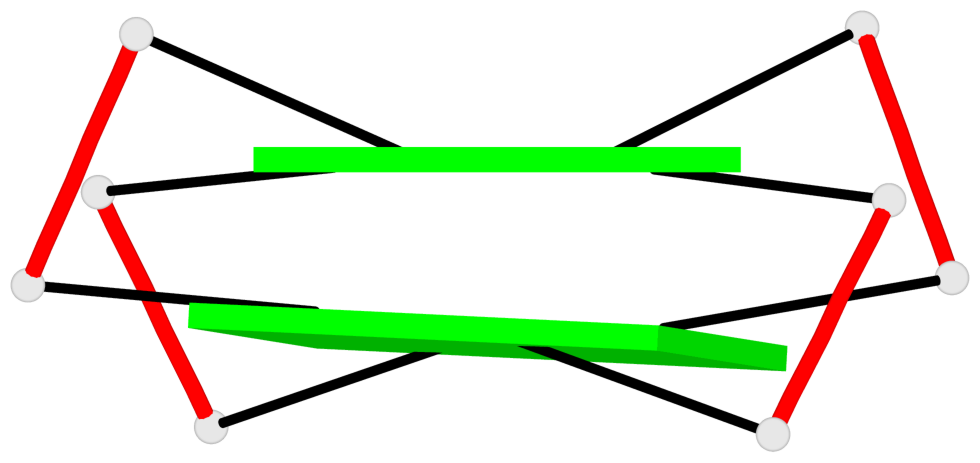 |
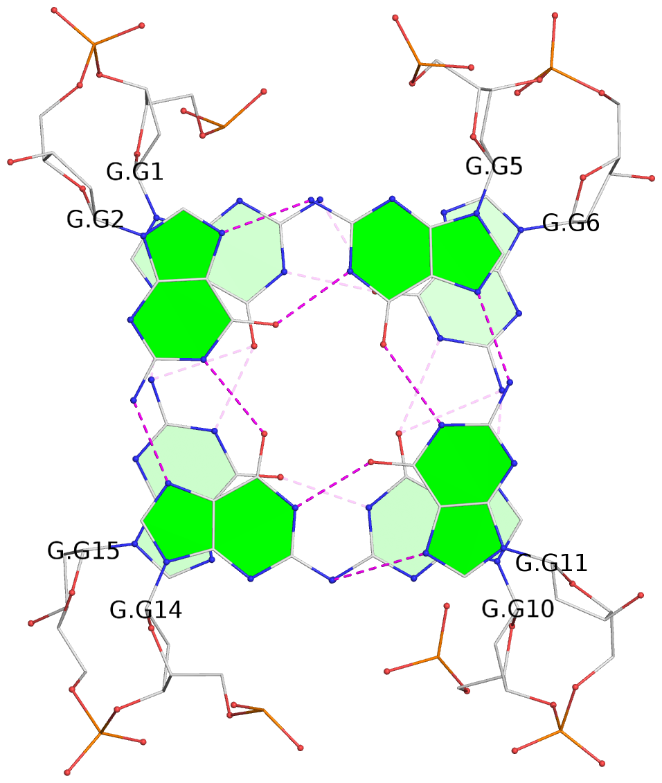
1 glyco-bond=s-s- sugar=-3-. groove=wnwn Major-->WC nts=4 GGGG G.DG1,G.DG15,G.DG10,G.DG6 2 glyco-bond=-s-s sugar=.--- groove=wnwn WC-->Major nts=4 GGGG G.DG2,G.DG14,G.DG11,G.DG5 step#1 mm(<>,outward) area=19.88 rise=3.50 twist=15.4 strand#1 DNA glyco-bond=s- sugar=-. nts=2 GG G.DG1,G.DG2 strand#2 DNA glyco-bond=-s sugar=3- nts=2 GG G.DG15,G.DG14 strand#3 DNA glyco-bond=s- sugar=-- nts=2 GG G.DG10,G.DG11 strand#4 DNA glyco-bond=-s sugar=.- nts=2 GG G.DG6,G.DG5 |
| 1 stacking diagram | |
 |
1 glyco-bond=s-s- sugar=-3-. groove=wnwn Major-->WC nts=4 GGGG G.DG1,G.DG15,G.DG10,G.DG6 |
Helix#4, 2 G-tetrad layers, INTRA-molecular, with 1 stem
 |
1 glyco-bond=s-s- sugar=-3-- groove=wnwn Major-->WC nts=4 GGGG I.DG1,I.DG15,I.DG10,I.DG6 2 glyco-bond=-s-s sugar=.-.- groove=wnwn WC-->Major nts=4 GGGG I.DG2,I.DG14,I.DG11,I.DG5 step#1 mm(<>,outward) area=15.91 rise=3.38 twist=16.6 strand#1 DNA glyco-bond=s- sugar=-. nts=2 GG I.DG1,I.DG2 strand#2 DNA glyco-bond=-s sugar=3- nts=2 GG I.DG15,I.DG14 strand#3 DNA glyco-bond=s- sugar=-. nts=2 GG I.DG10,I.DG11 strand#4 DNA glyco-bond=-s sugar=-- nts=2 GG I.DG6,I.DG5 |
| 1 stacking diagram | |
 |
1 glyco-bond=s-s- sugar=-3-- groove=wnwn Major-->WC nts=4 GGGG I.DG1,I.DG15,I.DG10,I.DG6 |
List of 4 G4-stems
In DSSR, a G4-stem is defined as a G4-helix with backbone connectivity. Bulges are also allowed along each of the four strands.
Stem#1, 2 G-tetrad layers, 3 loops, INTRA-molecular, UDUD, anti-parallel, 2(+Ln+Lw+Ln), chair(2+2)
 |
1 glyco-bond=s-s- sugar=---- groove=wnwn Major-->WC nts=4 GGGG E.DG1,E.DG15,E.DG10,E.DG6 2 glyco-bond=-s-s sugar=---- groove=wnwn WC-->Major nts=4 GGGG E.DG2,E.DG14,E.DG11,E.DG5 step#1 mm(<>,outward) area=19.44 rise=3.59 twist=14.7 strand#1 U DNA glyco-bond=s- sugar=-- nts=2 GG E.DG1,E.DG2 strand#2 D DNA glyco-bond=-s sugar=-- nts=2 GG E.DG15,E.DG14 strand#3 U DNA glyco-bond=s- sugar=-- nts=2 GG E.DG10,E.DG11 strand#4 D DNA glyco-bond=-s sugar=-- nts=2 GG E.DG6,E.DG5 loop#1 type=lateral strands=[#1,#4] nts=2 TT E.DT3,E.DT4 loop#2 type=lateral strands=[#4,#3] nts=3 TGT E.DT7,E.DG8,E.DT9 loop#3 type=lateral strands=[#3,#2] nts=2 TT E.DT12,E.DT13 |
Stem#2, 2 G-tetrad layers, 2 loops, INTRA-molecular, UDUD, anti-parallel, 2(+Ln+Ln), (2+2)
 |
1 glyco-bond=s-s- sugar=-3-. groove=wnwn Major-->WC nts=4 GGGG F.DG1,F.DG15,F.DG10,F.DG6 2 glyco-bond=-s-s sugar=--.- groove=wnwn WC-->Major nts=4 GGGG F.DG2,F.DG14,F.DG11,F.DG5 step#1 mm(<>,outward) area=16.82 rise=3.40 twist=16.9 strand#1 U DNA glyco-bond=s- sugar=-- nts=2 GG F.DG1,F.DG2 strand#2 D DNA glyco-bond=-s sugar=3- nts=2 GG F.DG15,F.DG14 strand#3 U DNA glyco-bond=s- sugar=-. nts=2 GG F.DG10,F.DG11 strand#4 D DNA glyco-bond=-s sugar=.- nts=2 GG F.DG6,F.DG5 loop#1 type=lateral strands=[#1,#4] nts=2 TT F.DT3,F.DT4 loop#2 type=lateral strands=[#3,#2] nts=2 TT F.DT12,F.DT13 |
Stem#3, 2 G-tetrad layers, 2 loops, INTRA-molecular, UDUD, anti-parallel, 2(+Ln+Ln), (2+2)
 |
1 glyco-bond=s-s- sugar=-3-. groove=wnwn Major-->WC nts=4 GGGG G.DG1,G.DG15,G.DG10,G.DG6 2 glyco-bond=-s-s sugar=.--- groove=wnwn WC-->Major nts=4 GGGG G.DG2,G.DG14,G.DG11,G.DG5 step#1 mm(<>,outward) area=19.88 rise=3.50 twist=15.4 strand#1 U DNA glyco-bond=s- sugar=-. nts=2 GG G.DG1,G.DG2 strand#2 D DNA glyco-bond=-s sugar=3- nts=2 GG G.DG15,G.DG14 strand#3 U DNA glyco-bond=s- sugar=-- nts=2 GG G.DG10,G.DG11 strand#4 D DNA glyco-bond=-s sugar=.- nts=2 GG G.DG6,G.DG5 loop#1 type=lateral strands=[#1,#4] nts=2 TT G.DT3,G.DT4 loop#2 type=lateral strands=[#3,#2] nts=2 TT G.DT12,G.DT13 |
Stem#4, 2 G-tetrad layers, 2 loops, INTRA-molecular, UDUD, anti-parallel, 2(+Ln+Ln), (2+2)
 |
1 glyco-bond=s-s- sugar=-3-- groove=wnwn Major-->WC nts=4 GGGG I.DG1,I.DG15,I.DG10,I.DG6 2 glyco-bond=-s-s sugar=.-.- groove=wnwn WC-->Major nts=4 GGGG I.DG2,I.DG14,I.DG11,I.DG5 step#1 mm(<>,outward) area=15.91 rise=3.38 twist=16.6 strand#1 U DNA glyco-bond=s- sugar=-. nts=2 GG I.DG1,I.DG2 strand#2 D DNA glyco-bond=-s sugar=3- nts=2 GG I.DG15,I.DG14 strand#3 U DNA glyco-bond=s- sugar=-. nts=2 GG I.DG10,I.DG11 strand#4 D DNA glyco-bond=-s sugar=-- nts=2 GG I.DG6,I.DG5 loop#1 type=lateral strands=[#1,#4] nts=2 TT I.DT3,I.DT4 loop#2 type=lateral strands=[#3,#2] nts=2 TT I.DT12,I.DT13 |