Detailed DSSR results for the G-quadruplex: PDB entry 8edp
Created and maintained by Xiang-Jun Lu <xiangjun@x3dna.org>
Citation: Please cite the NAR'20 DSSR-PyMOL schematics paper and/or the NAR'15 DSSR method paper.
Summary information
- PDB id
- 8edp
- Class
- DNA
- Method
- X-ray (2.01 Å)
- Summary
- Crystal structure of a three-tetrad, parallel, and k+ stabilized homopurine g-quadruplex from human chromosome 7
- Reference
- Ye M, Chen EV, Pfeil SH, Martin KN, Atrafi T, Yun S, Martinez Z, Yatsunyk LA (2022): "Homopurine guanine-rich sequences in complex with N-methyl mesoporphyrin IX form parallel G-quadruplex dimers and display a unique symmetry tetrad." Bioorg.Med.Chem., 77, 117112. doi: 10.1016/j.bmc.2022.117112.
- Abstract
- DNA can fold into G-quadruplexes (GQs), non-canonical secondary structures formed by π-π stacking of G-tetrads. GQs are important in many biological processes, which makes them promising therapeutic targets. We identified a 42-nucleotide long, purine-only G-rich sequence from human genome, which contains eight G-stretches connected by A and AAAA loops. We divided this sequence into five unique segments, four guanine stretches each, named GA1-5. In order to investigate the role of adenines in GQ structure formation, we performed biophysical and X-ray crystallographic studies of GA1-5 and their complexes with a highly selective GQ ligand, N-methyl mesoporphyrin IX (NMM). Our data indicate that all variants form parallel GQs whose stability depends on the number of flexible AAAA loops. GA1-3 bind NMM with 1:1 stoichiometry. The Ka for GA1 and GA3 is modest, ∼0.3 μM -1, and that for GA2 is significantly higher, ∼1.2 μM -1. NMM stabilizes GA1-3 by 14.6, 13.1, and 7.0 ℃, respectively, at 2 equivalents. We determined X-ray crystal structures of GA1-NMM (1.98 Å resolution) and GA3-NMM (2.01 Å). The structures confirm the parallel topology of GQs with all adenines forming loops and display NMM binding at the 3' G-tetrad. Both complexes dimerize through the 5' interface. We observe two novel structural features: 1) a 'symmetry tetrad' at the dimer interface, which is formed by two guanines from each GQ monomer and 2) a NMM dimer in GA1-NMM. Our structural work confirms great flexibility of adenines as structural elements in GQ formation and contributes greatly to our understanding of the structural diversity of GQs and their modes of interaction with small molecule ligands.
- G4 notes
- 3 G-tetrads, 1 G4 helix, 1 G4 stem, 3(-P-P-P), parallel(4+0), UUUU
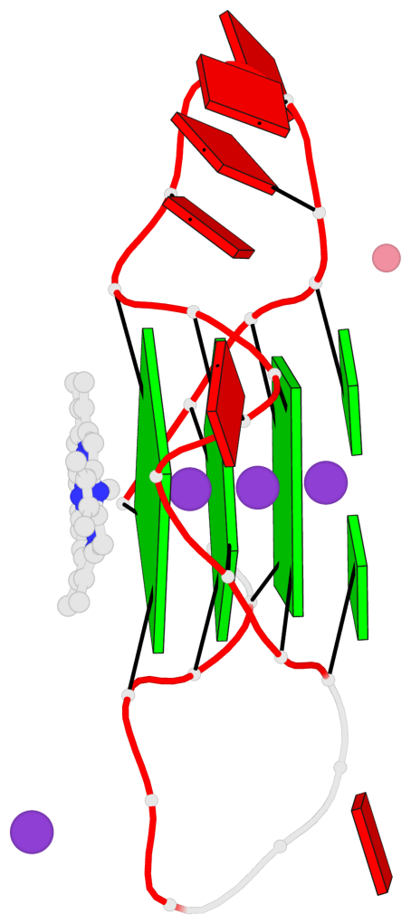
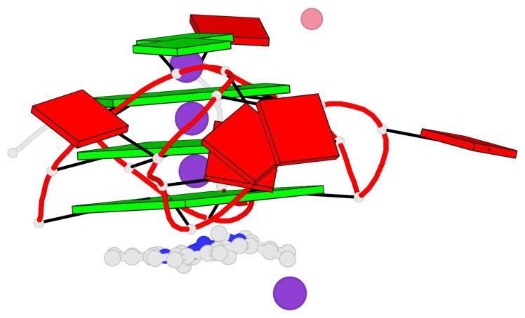
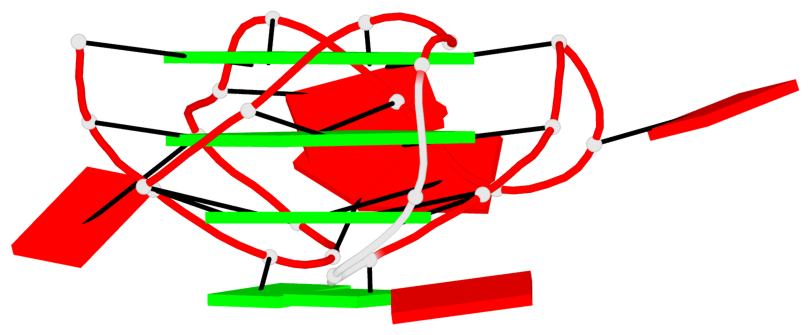
Base-block schematics in six views
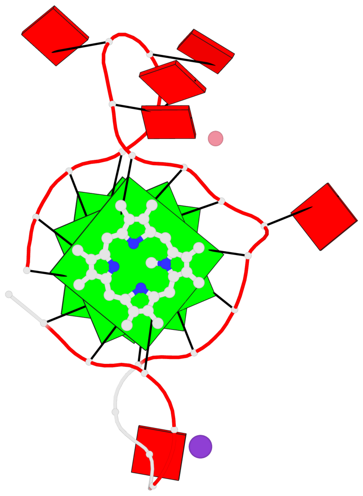
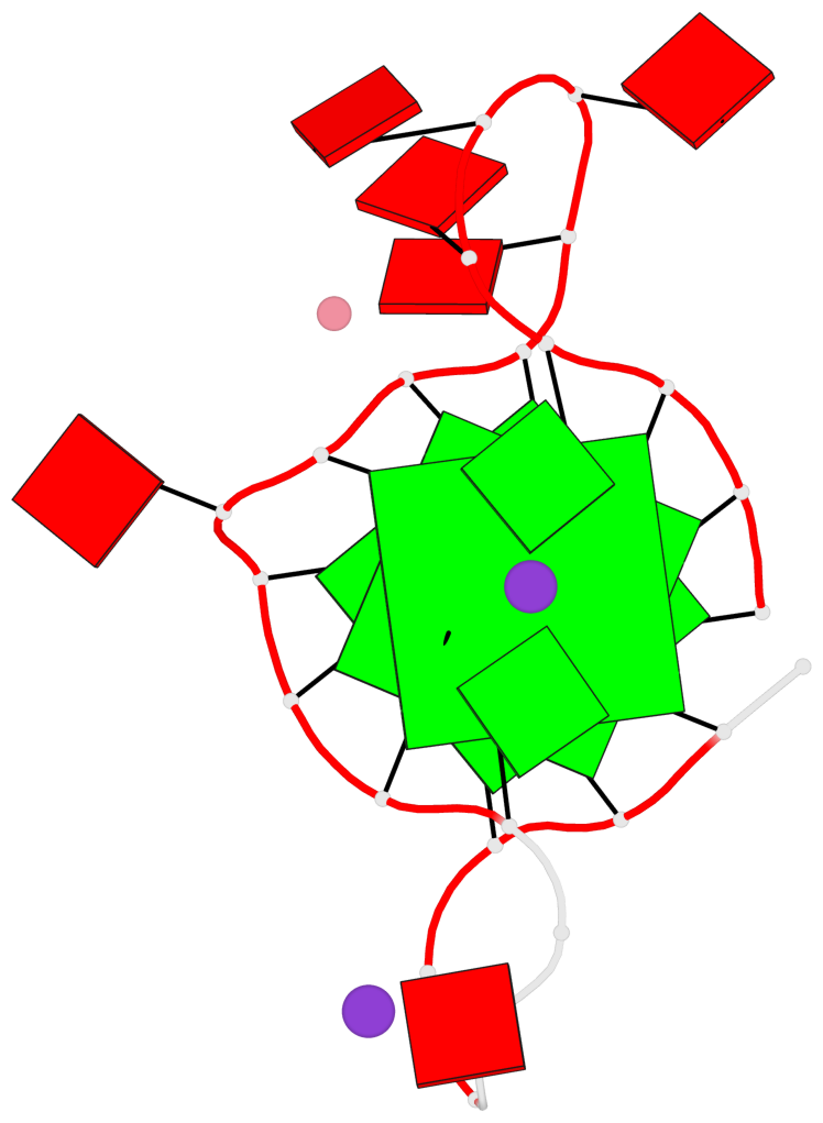
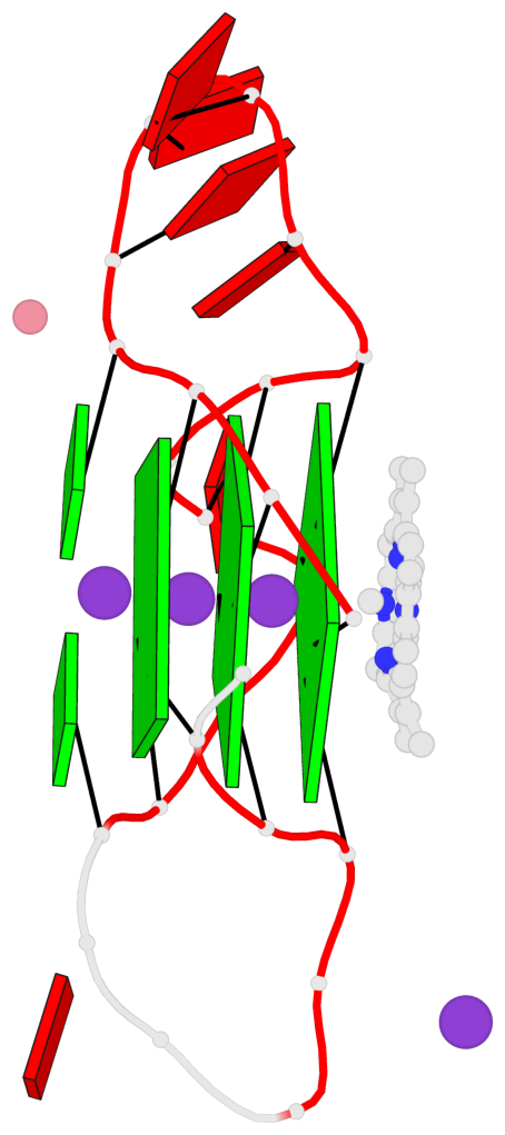
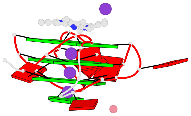
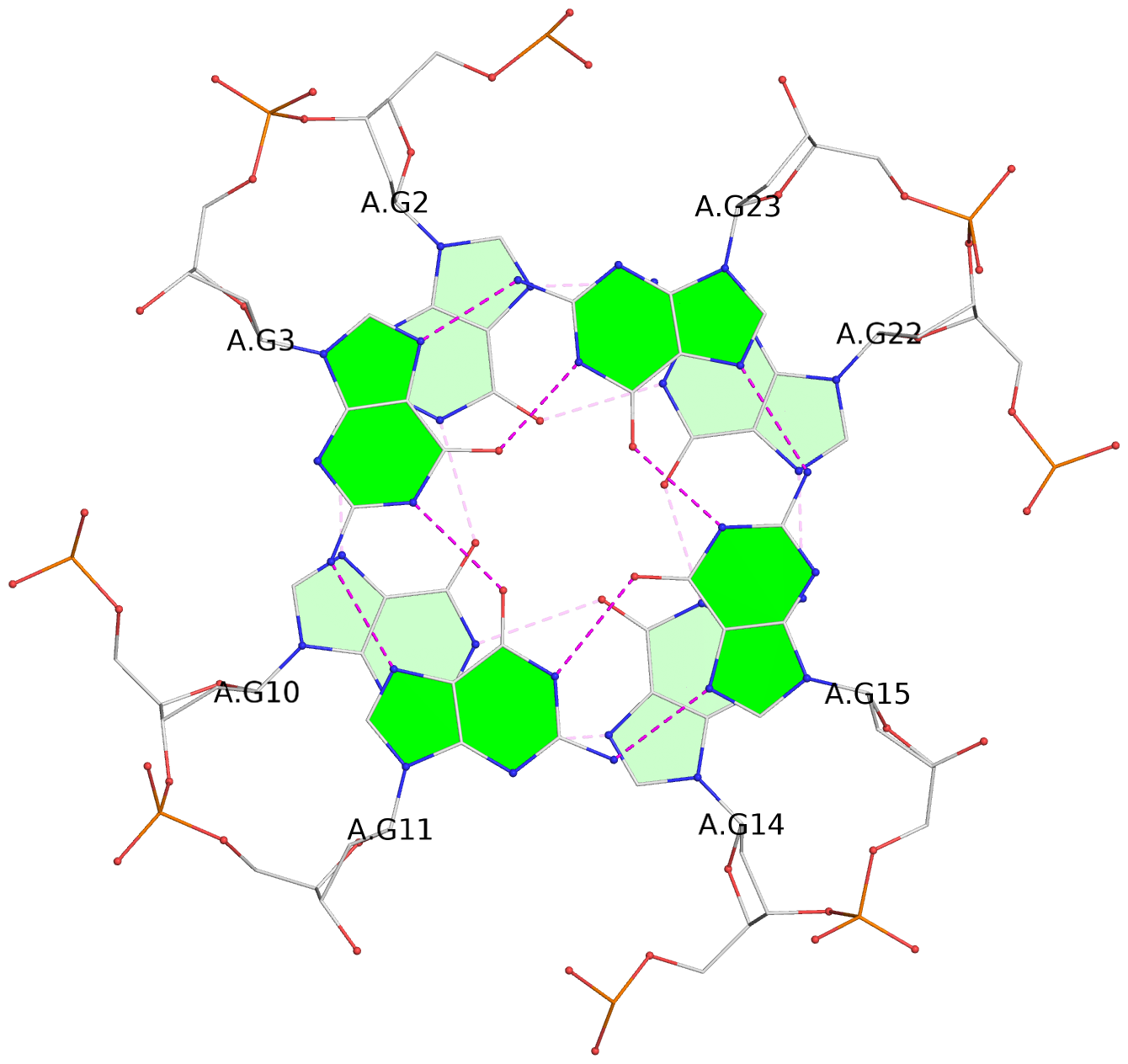
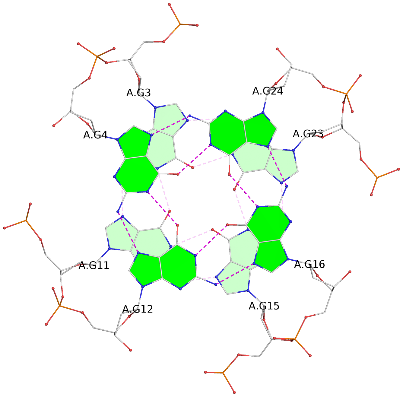
List of 3 G-tetrads
 1 glyco-bond=---- sugar=---- groove=---- planarity=0.182 type=bowl-2 nts=4 GGGG A.DG2,A.DG10,A.DG14,A.DG22
 2 glyco-bond=---- sugar=---- groove=---- planarity=0.227 type=bowl-2 nts=4 GGGG A.DG3,A.DG11,A.DG15,A.DG23
 3 glyco-bond=---- sugar=---- groove=---- planarity=0.324 type=bowl   nts=4 GGGG A.DG4,A.DG12,A.DG16,A.DG24
List of 1 G4-helix
In DSSR, a G4-helix is defined by stacking interactions of G-tetrads, regardless of backbone connectivity, and may contain more than one G4-stem.
Helix#1, 3 G-tetrad layers, INTRA-molecular, with 1 stem
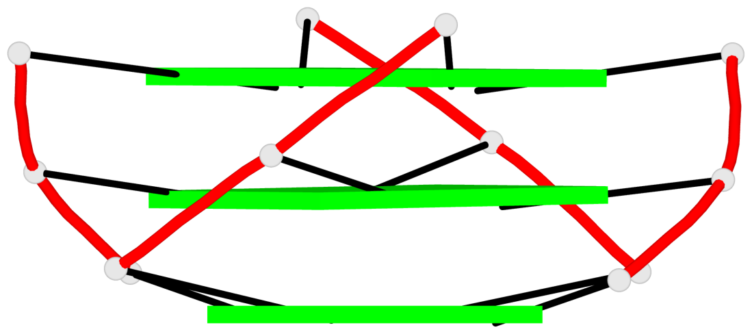 |
1 glyco-bond=---- sugar=---- groove=---- WC-->Major nts=4 GGGG A.DG2,A.DG10,A.DG14,A.DG22 2 glyco-bond=---- sugar=---- groove=---- WC-->Major nts=4 GGGG A.DG3,A.DG11,A.DG15,A.DG23 3 glyco-bond=---- sugar=---- groove=---- WC-->Major nts=4 GGGG A.DG4,A.DG12,A.DG16,A.DG24 step#1 pm(>>,forward) area=10.78 rise=3.36 twist=30.7 step#2 pm(>>,forward) area=11.16 rise=3.43 twist=27.8 strand#1 DNA glyco-bond=--- sugar=--- nts=3 GGG A.DG2,A.DG3,A.DG4 strand#2 DNA glyco-bond=--- sugar=--- nts=3 GGG A.DG10,A.DG11,A.DG12 strand#3 DNA glyco-bond=--- sugar=--- nts=3 GGG A.DG14,A.DG15,A.DG16 strand#4 DNA glyco-bond=--- sugar=--- nts=3 GGG A.DG22,A.DG23,A.DG24 |
| 2 stacking diagrams | |
 |
1 glyco-bond=---- sugar=---- groove=---- WC-->Major nts=4 GGGG A.DG2,A.DG10,A.DG14,A.DG22 |
 |
2 glyco-bond=---- sugar=---- groove=---- WC-->Major nts=4 GGGG A.DG3,A.DG11,A.DG15,A.DG23 |
List of 1 G4-stem
In DSSR, a G4-stem is defined as a G4-helix with backbone connectivity. Bulges are also allowed along each of the four strands.
Stem#1, 3 G-tetrad layers, 3 loops, INTRA-molecular, UUUU, parallel, 3(-P-P-P), parallel(4+0)
 |
1 glyco-bond=---- sugar=---- groove=---- WC-->Major nts=4 GGGG A.DG2,A.DG10,A.DG14,A.DG22 2 glyco-bond=---- sugar=---- groove=---- WC-->Major nts=4 GGGG A.DG3,A.DG11,A.DG15,A.DG23 3 glyco-bond=---- sugar=---- groove=---- WC-->Major nts=4 GGGG A.DG4,A.DG12,A.DG16,A.DG24 step#1 pm(>>,forward) area=10.78 rise=3.36 twist=30.7 step#2 pm(>>,forward) area=11.16 rise=3.43 twist=27.8 strand#1 U DNA glyco-bond=--- sugar=--- nts=3 GGG A.DG2,A.DG3,A.DG4 strand#2 U DNA glyco-bond=--- sugar=--- nts=3 GGG A.DG10,A.DG11,A.DG12 strand#3 U DNA glyco-bond=--- sugar=--- nts=3 GGG A.DG14,A.DG15,A.DG16 strand#4 U DNA glyco-bond=--- sugar=--- nts=3 GGG A.DG22,A.DG23,A.DG24 loop#1 type=propeller strands=[#1,#2] nts=5 AAAAG A.DA5,A.DA6,A.DA7,A.DA8,A.DG9 loop#2 type=propeller strands=[#2,#3] nts=1 A A.DA13 loop#3 type=propeller strands=[#3,#4] nts=5 AAAAG A.DA17,A.DA18,A.DA19,A.DA20,A.DG21 |