Detailed DSSR results for the G-quadruplex: PDB entry 8psc
Created and maintained by Xiang-Jun Lu <xiangjun@x3dna.org>
Citation: Please cite the NAR'20 DSSR-PyMOL schematics paper and/or the NAR'15 DSSR method paper.
Summary information
- PDB id
- 8psc
- Class
- DNA
- Method
- NMR
- Summary
- Three-layered basket-type g-quadruplex from a g-rich sequence with five g-runs
- Reference
- Vianney YM, Schroder N, Jana J, Chojetzki G, Weisz K (2023): "Showcasing Different G-Quadruplex Folds of a G-Rich Sequence: Between Rule-Based Prediction and Butterfly Effect." J.Am.Chem.Soc., 145, 22194-22205. doi: 10.1021/jacs.3c08336.
- Abstract
- In better understanding the interactions of G-quadruplexes in a cellular or noncellular environment, a reliable sequence-based prediction of their three-dimensional fold would be extremely useful, yet is often limited by their remarkable structural diversity. A G-rich sequence related to a promoter sequence of the PDGFR-β nuclease hypersensitivity element (NHE) comprises a G3-G3-G2-G4-G3 pattern of five G-runs with two to four G residues. Although the predominant formation of three-layered canonical G-quadruplexes with uninterrupted G-columns can be expected, minimal base substitutions in a non-G-tract domain were shown to guide folding into either a basket-type antiparallel quadruplex, a parallel-stranded quadruplex with an interrupted G-column, a quadruplex with a V-shaped loop, or a (3+1) hybrid quadruplex. A 3D NMR structure for each of the different folds was determined. Supported by thermodynamic profiling on additional sequence variants, formed topologies were rationalized by the identification and assessment of specific critical interactions of loop and overhang residues, giving valuable insights into their contribution to favor a particular conformer. The variability of such tertiary interactions, together with only small differences in quadruplex free energies, emphasizes current limits for a reliable sequence-dependent prediction of favored topologies from sequences with multiple irregularly positioned G-tracts.
- G4 notes
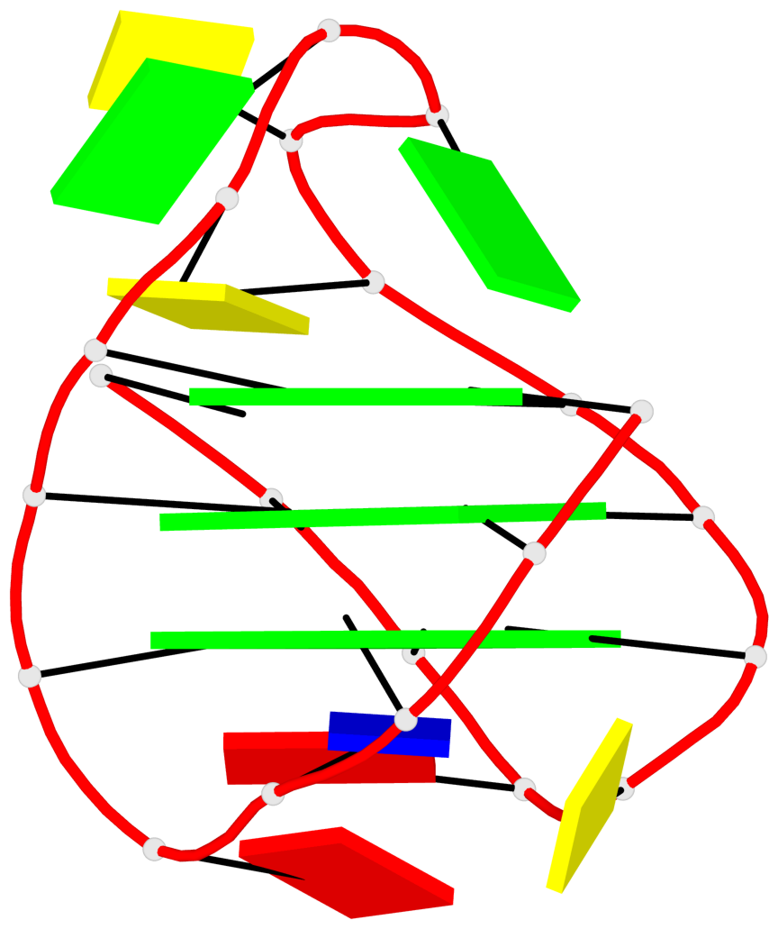
- 3 G-tetrads, 1 G4 helix, 1 G4 stem, 3(-LwD+Ln), basket(2+2), UDDU
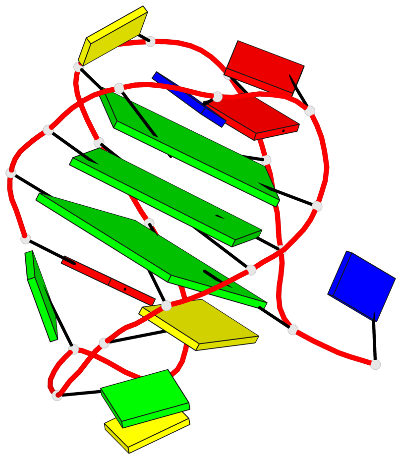
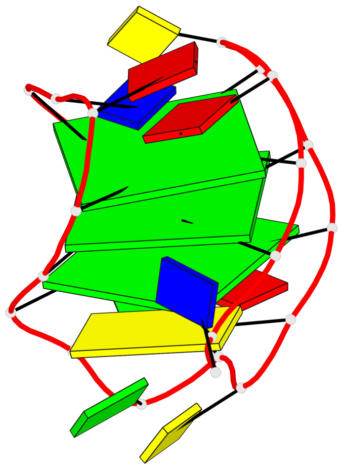
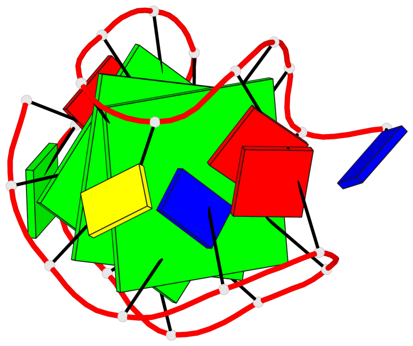
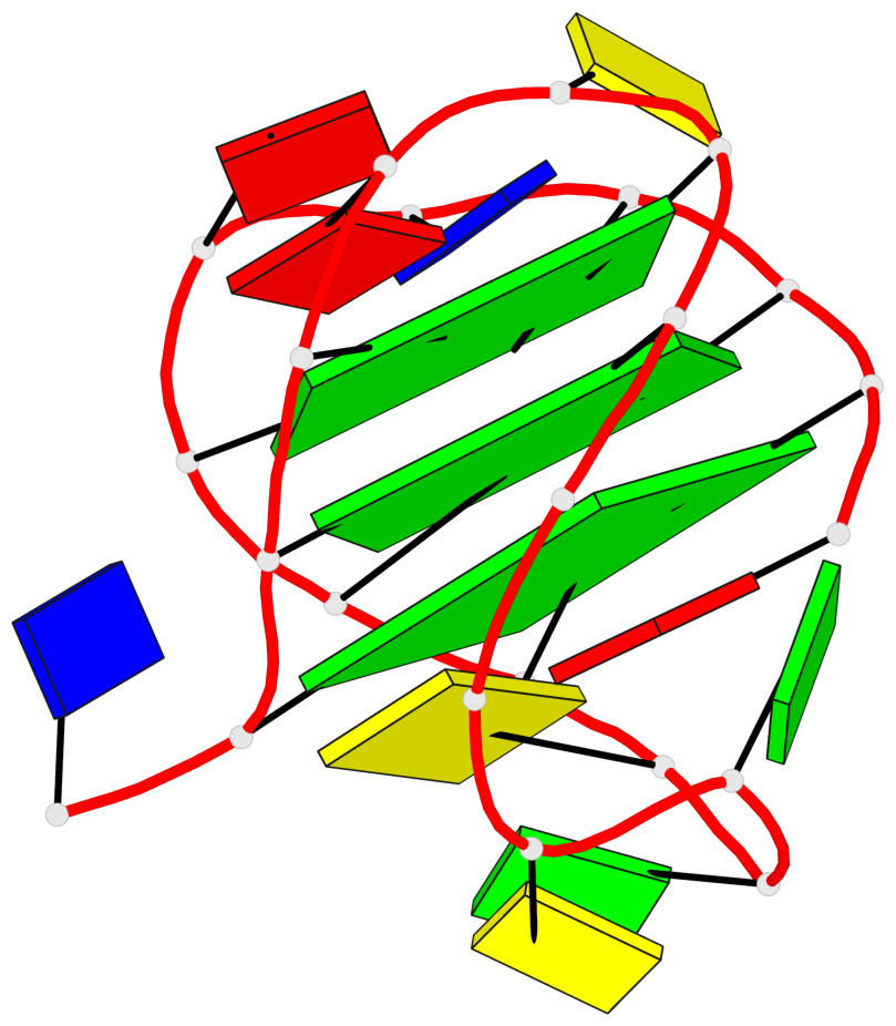
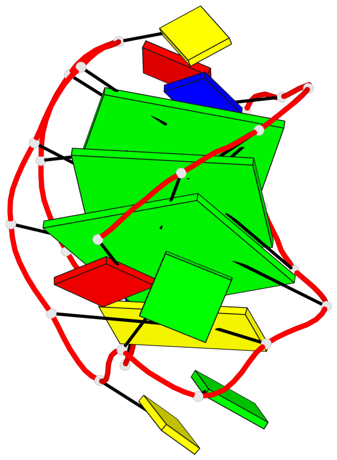
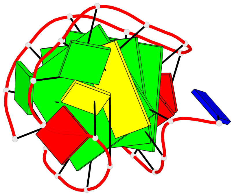
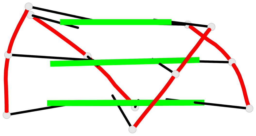
Base-block schematics in six views
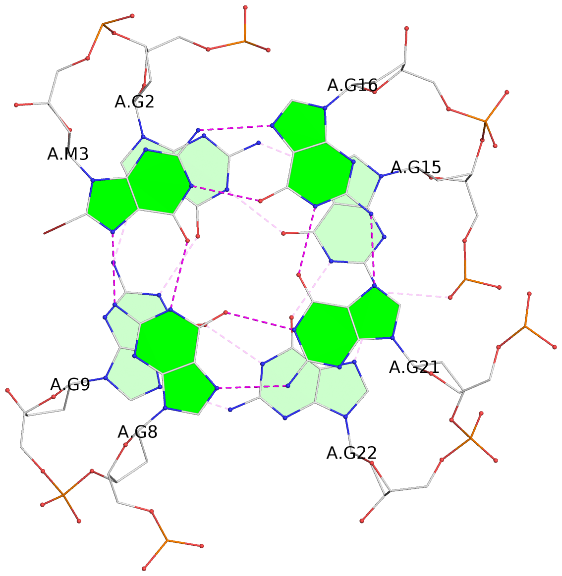
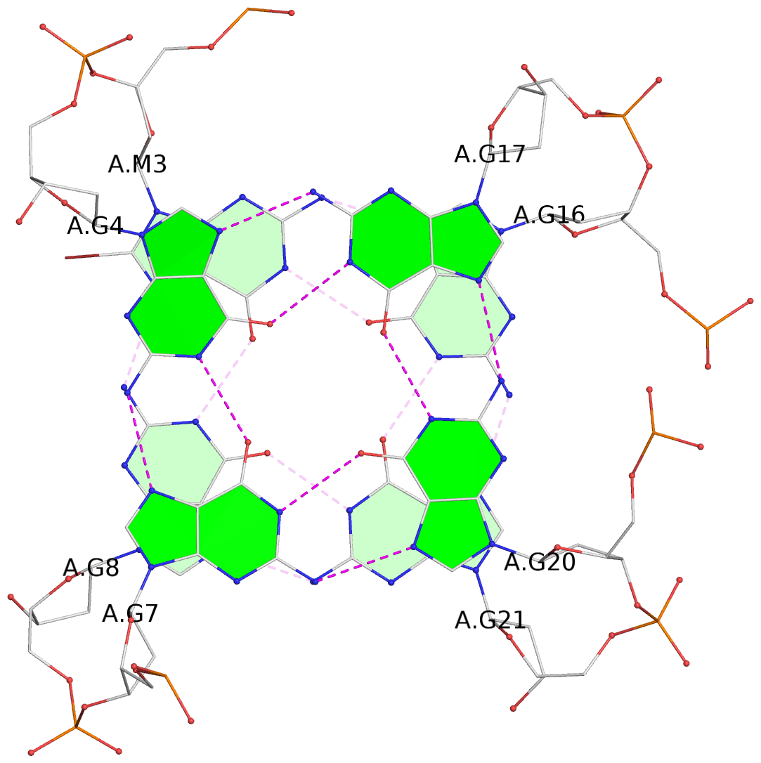
List of 3 G-tetrads
 1 glyco-bond=s--s sugar=---- groove=w-n- planarity=0.174 type=other  nts=4 GGGG A.DG2,A.DG9,A.DG22,A.DG15
 2 glyco-bond=s--s sugar=---- groove=w-n- planarity=0.176 type=other  nts=4 gGGG A.BGM3,A.DG8,A.DG21,A.DG16
 3 glyco-bond=-ss- sugar=---- groove=w-n- planarity=0.338 type=bowl   nts=4 GGGG A.DG4,A.DG7,A.DG20,A.DG17
List of 1 G4-helix
In DSSR, a G4-helix is defined by stacking interactions of G-tetrads, regardless of backbone connectivity, and may contain more than one G4-stem.
Helix#1, 3 G-tetrad layers, INTRA-molecular, with 1 stem
 |
1 glyco-bond=s--s sugar=---- groove=w-n- Major-->WC nts=4 GGGG A.DG2,A.DG9,A.DG22,A.DG15 2 glyco-bond=s--s sugar=---- groove=w-n- Major-->WC nts=4 gGGG A.BGM3,A.DG8,A.DG21,A.DG16 3 glyco-bond=-ss- sugar=---- groove=w-n- WC-->Major nts=4 GGGG A.DG4,A.DG7,A.DG20,A.DG17 step#1 mp(<<,backward) area=13.13 rise=3.46 twist=26.3 step#2 mm(<>,outward) area=18.17 rise=3.55 twist=14.9 strand#1 DNA glyco-bond=ss- sugar=--- nts=3 GgG A.DG2,A.BGM3,A.DG4 strand#2 DNA glyco-bond=--s sugar=--- nts=3 GGG A.DG9,A.DG8,A.DG7 strand#3 DNA glyco-bond=--s sugar=--- nts=3 GGG A.DG22,A.DG21,A.DG20 strand#4 DNA glyco-bond=ss- sugar=--- nts=3 GGG A.DG15,A.DG16,A.DG17 |
| 2 stacking diagrams | |
 |
1 glyco-bond=s--s sugar=---- groove=w-n- Major-->WC nts=4 GGGG A.DG2,A.DG9,A.DG22,A.DG15 |
 |
2 glyco-bond=s--s sugar=---- groove=w-n- Major-->WC nts=4 gGGG A.BGM3,A.DG8,A.DG21,A.DG16 |
List of 1 G4-stem
In DSSR, a G4-stem is defined as a G4-helix with backbone connectivity. Bulges are also allowed along each of the four strands.
Stem#1, 3 G-tetrad layers, 3 loops, INTRA-molecular, UDDU, anti-parallel, 3(-LwD+Ln), basket(2+2)
 |
1 glyco-bond=s--s sugar=---- groove=w-n- Major-->WC nts=4 GGGG A.DG2,A.DG9,A.DG22,A.DG15 2 glyco-bond=s--s sugar=---- groove=w-n- Major-->WC nts=4 gGGG A.BGM3,A.DG8,A.DG21,A.DG16 3 glyco-bond=-ss- sugar=---- groove=w-n- WC-->Major nts=4 GGGG A.DG4,A.DG7,A.DG20,A.DG17 step#1 mp(<<,backward) area=13.13 rise=3.46 twist=26.3 step#2 mm(<>,outward) area=18.17 rise=3.55 twist=14.9 strand#1 U DNA glyco-bond=ss- sugar=--- nts=3 GgG A.DG2,A.BGM3,A.DG4 strand#2 D DNA glyco-bond=--s sugar=--- nts=3 GGG A.DG9,A.DG8,A.DG7 strand#3 D DNA glyco-bond=--s sugar=--- nts=3 GGG A.DG22,A.DG21,A.DG20 strand#4 U DNA glyco-bond=ss- sugar=--- nts=3 GGG A.DG15,A.DG16,A.DG17 loop#1 type=lateral strands=[#1,#2] nts=2 TA A.DT5,A.DA6 loop#2 type=diagonal strands=[#2,#4] nts=5 CGGCG A.DC10,A.DG11,A.DG12,A.DC13,A.DG14 loop#3 type=lateral strands=[#4,#3] nts=2 CA A.DC18,A.DA19 |