Detailed DSSR results for the G-quadruplex: PDB entry 1a6h
Created and maintained by Xiang-Jun Lu <xiangjun@x3dna.org>
Citation: Please cite the NAR'20 DSSR-PyMOL schematics paper and/or the NAR'15 DSSR method paper.
Summary information
- PDB id
- 1a6h
- Class
- DNA
- Method
- NMR
- Summary
- DNA quadruplex containing gcgc tetrad, NMR, 4 structures
- Reference
- Kettani A, Kumar RA, Patel DJ (1995): "Solution structure of a DNA quadruplex containing the fragile X syndrome triplet repeat." J.Mol.Biol., 254, 638-656. doi: 10.1006/jmbi.1995.0644.
- Abstract
- Both X-ray and NMR structural studies have defined the polymorphic nature of G-quadruplexes generated through mutual stacking of G.G.G.G tetrads by guanine rich telomeric sequences. Recently, the fragile X syndrome d(C-G-G)n triplet nucleotide repeat has been shown to form a stable quadruplex of undefined structure in monovalent cation solution. We have undertaken a structural characterization of the d(G-C-G-G-T3-G-C-G-G) undecanucleotide to elucidate the structural alignments associated with quadruplex formation by this oligomer which contains sequence elements associated with the fragile X syndrome triplet repeat. d(G-C-G-G-T3-G-C-G-G) in Na+ cation solution forms a quadruplex through dimerization of two symmetry related hairpins with the lateral connecting T3 loops positioned at opposite ends of the quadruplex. This novel NMR-molecular dynamics based solution structure contains internal G.C.G.C tetrads sandwiched between terminal G.G.G.G tetrads. Watson-Crick G.C base-pairs within individual hairpins dimerize through their major groove edges using bifurcated hydrogen bonds to form internal G(anti).C(anti).G(anti).C(anti) tetrads. Adjacent strands are anti-parallel to each other around the symmetric G-quadruplex which contains two distinct narrow and two symmetric wide grooves. By contrast, the terminal G-tetrads adopt G(syn).G(anti).G(syn).G(anti) alignments. The structure of the d(G-C-G-G-T3-G-C-G-G) quadruplex with its multi-layer arrangement of G.G.G.G and G.C.G.C tetrads greatly expands on our current knowledge of quadruplex folding topologies. Our results establish the pairing alignments that can be potentially utilized by the fragile X syndrome triplet repeat to form quadruplex structures through dimerization of hairpin stems. The formation of novel G.C.G.C tetrads through dimerization of Watson-Crick G.C base-pairs is directly relevant to the potential pairing alignments of helical stems in genetic recombination.
- G4 notes
- 2 G-tetrads
Base-block schematics in six views
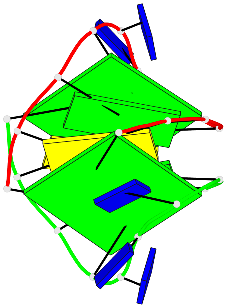
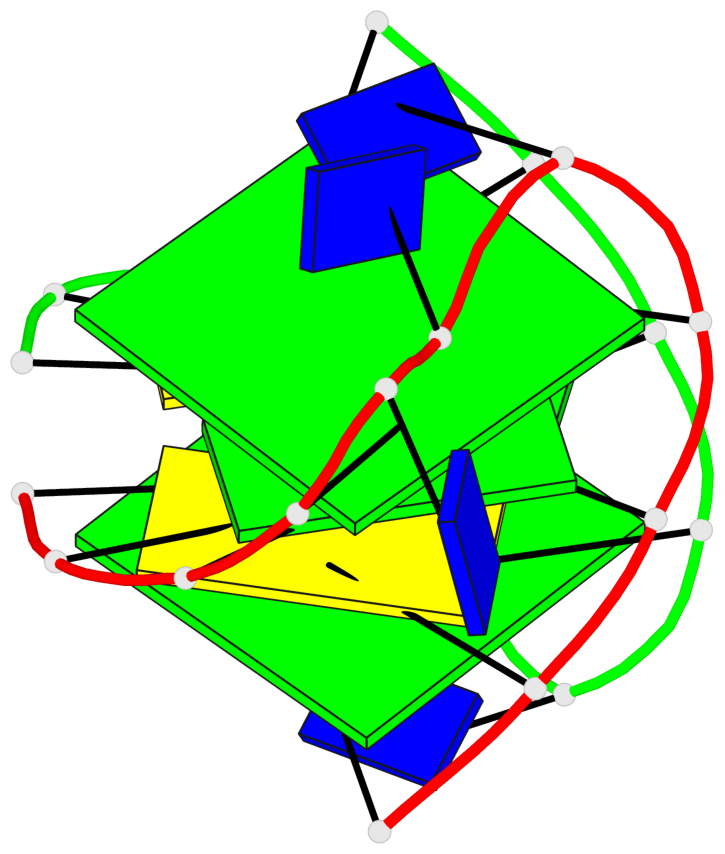
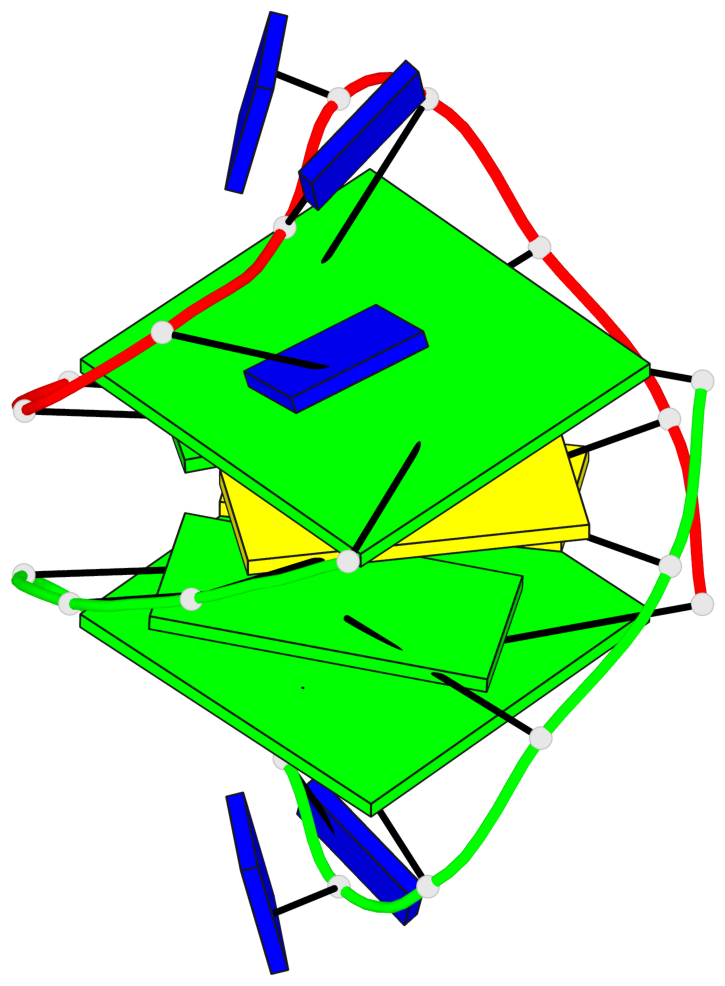
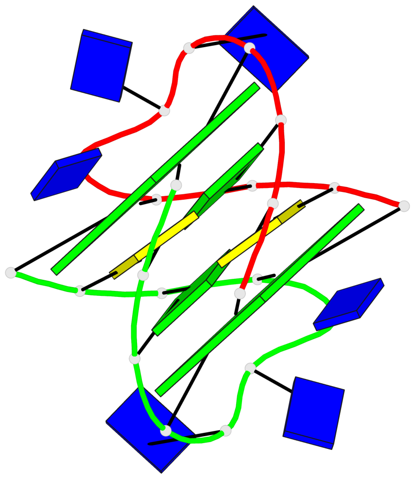
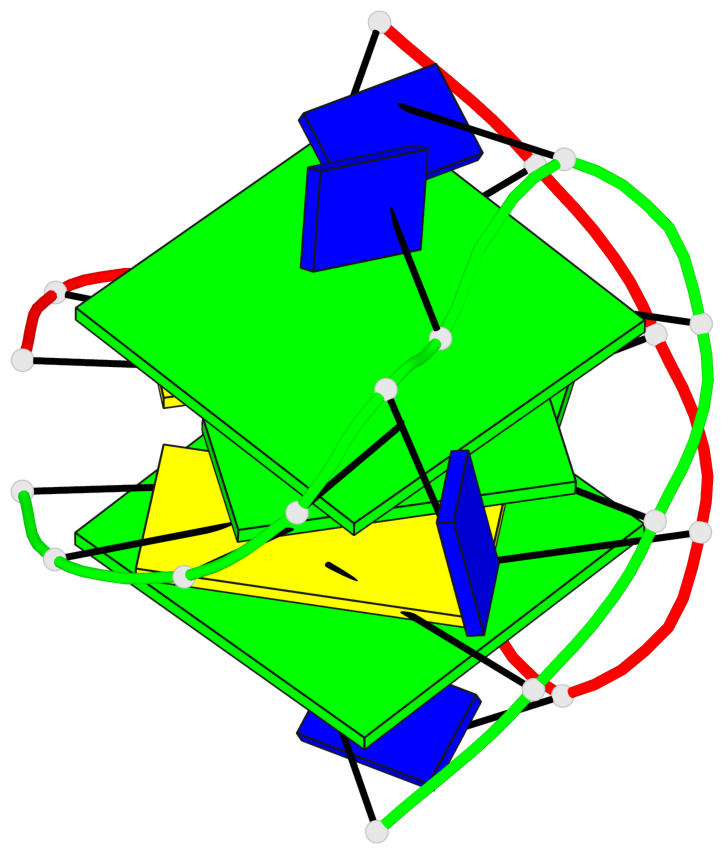
List of 2 G-tetrads
 1 glyco-bond=s-s- sugar=---. groove=wnwn planarity=0.390 type=bowl   nts=4 GGGG A.DG1,A.DG11,B.DG8,B.DG4
 2 glyco-bond=-s-s sugar=---- groove=wnwn planarity=0.391 type=bowl   nts=4 GGGG A.DG4,A.DG8,B.DG11,B.DG1
List of 2 non-stem G4-loops (including the two closing Gs)
 1 type=lateral   helix=#-2 nts=5 GTTTG A.DG4,A.DT5,A.DT6,A.DT7,A.DG8
 2 type=lateral   helix=#-1 nts=5 GTTTG B.DG4,B.DT5,B.DT6,B.DT7,B.DG8