Detailed DSSR results for the G-quadruplex: PDB entry 1a8w
Created and maintained by Xiang-Jun Lu <xiangjun@x3dna.org>
Citation: Please cite the NAR'20 DSSR-PyMOL schematics paper and/or the NAR'15 DSSR method paper.
Summary information
- PDB id
- 1a8w
- Class
- DNA
- Method
- NMR
- Summary
- A k+ cation-induced conformational switch within a loop spanning segment of a DNA quadruplex containing g-g-g-c repeats, NMR, 8 structures
- Reference
- Bouaziz S, Kettani A, Patel DJ (1998): "A K cation-induced conformational switch within a loop spanning segment of a DNA quadruplex containing G-G-G-C repeats." J.Mol.Biol., 282, 637-652. doi: 10.1006/jmbi.1998.2031.
- Abstract
- We have identified a unique structural transition (in slow exchange on the NMR time scale) in the tertiary fold of the d(G-G-G-C-T4-G-G-G-C) quadruplex on proceeding from Na+ to K+ as counterion in aqueous solution. Both monovalent cation-dependent conformations exhibit certain common structural features, which include head-to-tail dimerization of two symmetry-related stem-hairpin loops, adjacent strands which are antiparallel to each other and adjacent stacked G(syn).G(anti). G(syn).G(anti) tetrads in the central core of the quadruplexes. The Na and K cation stabilized structures of the d(G-G-G-C-T4-G-G-G-C) quadruplexes differ in the conformations of the T-T-T-T loops, the relative alignment of G.C base-pairs positioned opposite each other through their major groove edges and potentially in the number of monovalent cation binding sites. We have identified potential K cation binding cavities within the symmetry-related T-T-T-G segments, suggesting the potential for two additional monovalent cation binding sites in the K cation-stabilized quadruplex relative to its Na cation-stabilized counterpart. Modeling studies suggest that the major groove edges of guanine residues in Watson-Crick G.C base-pairs could potentially be bridged by coordinated K cations in the d(G-G-G-C-T4-G-G-G-C) quadruplex in KCl solution in contrast to formation of G.C.G.C tetrads for the corresponding quadruplex in NaCl solution. Our results defining the molecular basis of a Na to K cation-dependent conformational switch in the loop spanning segment of the d(G-G-G-C-T4-G-G-G-C) quadruplex may have relevance to recent observations that specific K cation coordinated loop conformations within quadruplexes exhibit inhibitory activity against HIV integrase.
- G4 notes
- 2 G-tetrads, 1 G4 helix, 1 G4 stem, (2+2), UDUD
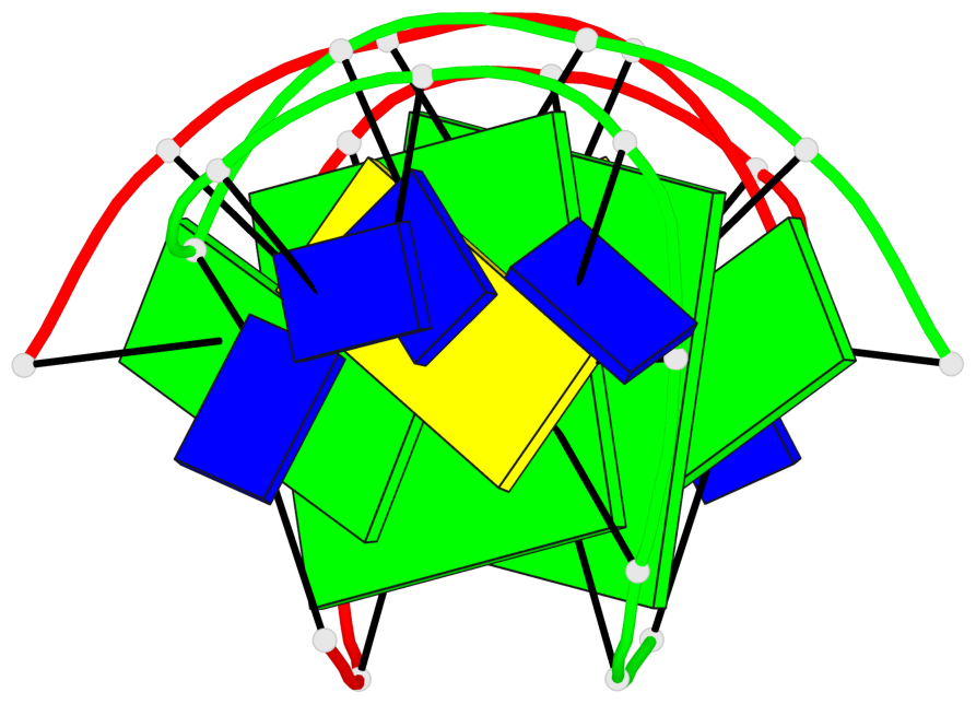
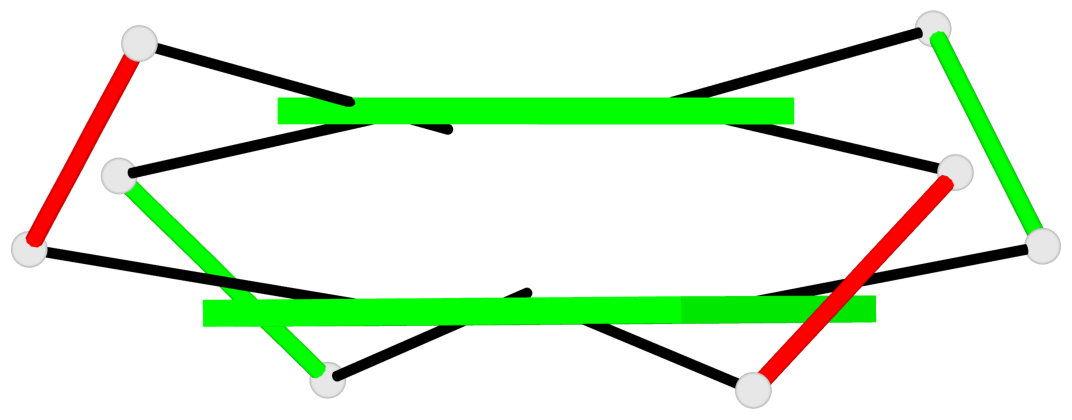
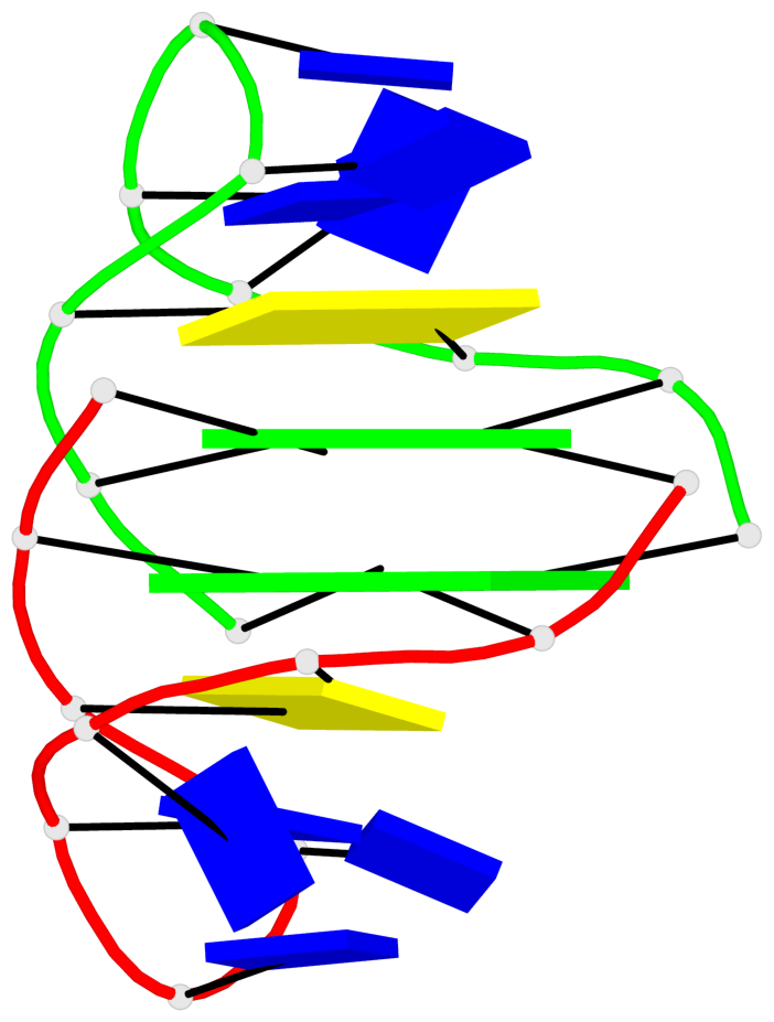
Base-block schematics in six views
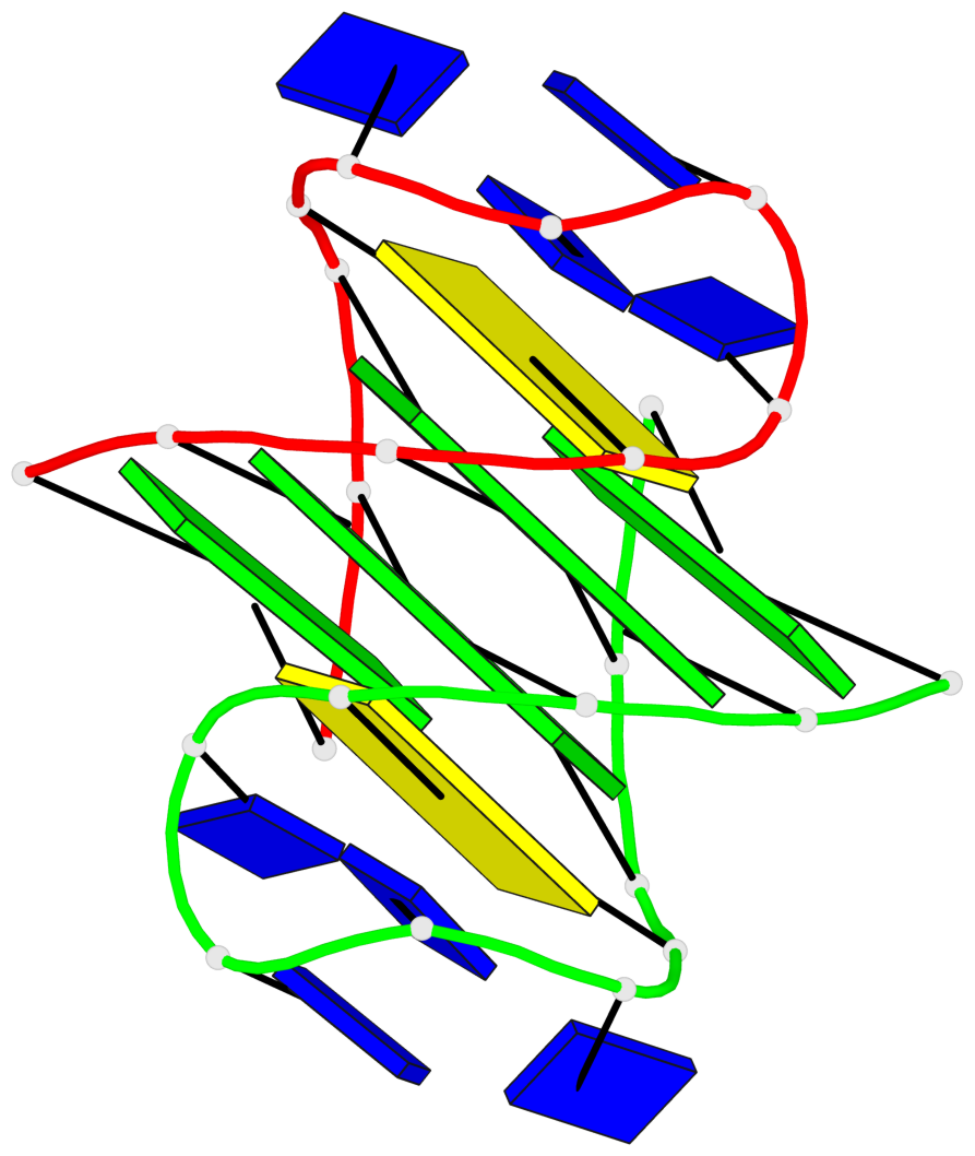
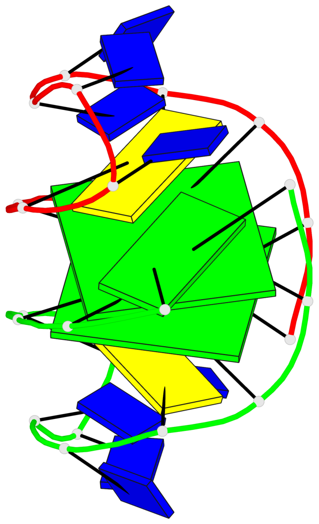
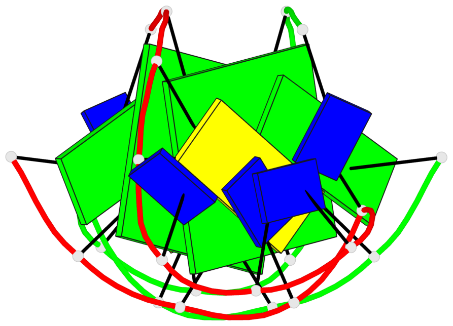
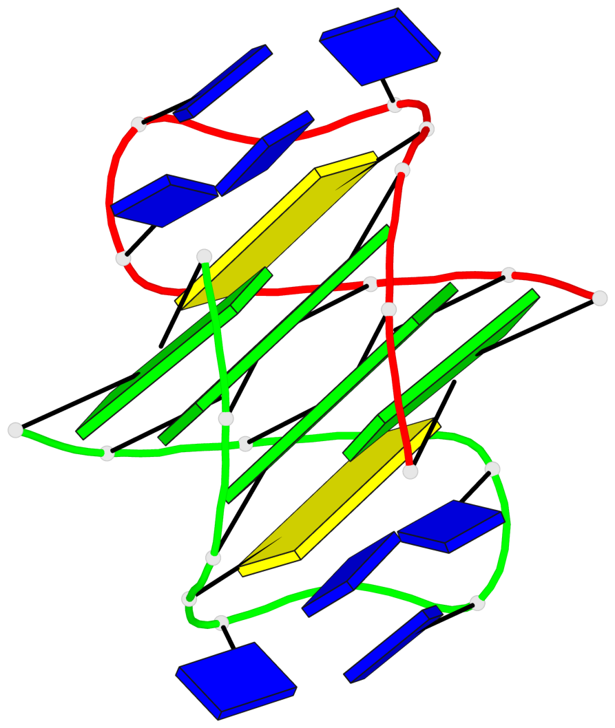
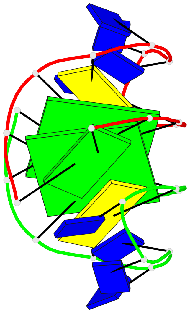
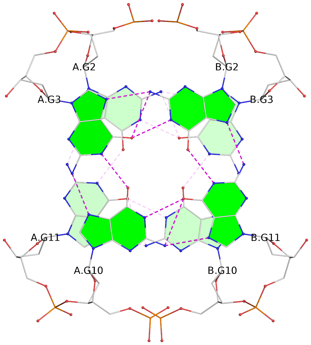
List of 2 G-tetrads
 1 glyco-bond=s-s- sugar=---- groove=wnwn planarity=0.383 type=bowl-2 nts=4 GGGG A.DG2,A.DG11,B.DG10,B.DG3
 2 glyco-bond=-s-s sugar=---- groove=wnwn planarity=0.382 type=bowl-2 nts=4 GGGG A.DG3,A.DG10,B.DG11,B.DG2
List of 1 G4-helix
In DSSR, a G4-helix is defined by stacking interactions of G-tetrads, regardless of backbone connectivity, and may contain more than one G4-stem.
Helix#1, 2 G-tetrad layers, inter-molecular, with 1 stem
 |
1 glyco-bond=s-s- sugar=---- groove=wnwn Major-->WC nts=4 GGGG A.DG2,A.DG11,B.DG10,B.DG3 2 glyco-bond=-s-s sugar=---- groove=wnwn WC-->Major nts=4 GGGG A.DG3,A.DG10,B.DG11,B.DG2 step#1 mm(<>,outward) area=14.60 rise=3.72 twist=22.1 strand#1 DNA glyco-bond=s- sugar=-- nts=2 GG A.DG2,A.DG3 strand#2 DNA glyco-bond=-s sugar=-- nts=2 GG A.DG11,A.DG10 strand#3 DNA glyco-bond=s- sugar=-- nts=2 GG B.DG10,B.DG11 strand#4 DNA glyco-bond=-s sugar=-- nts=2 GG B.DG3,B.DG2 |
| 1 stacking diagram | |
 |
1 glyco-bond=s-s- sugar=---- groove=wnwn Major-->WC nts=4 GGGG A.DG2,A.DG11,B.DG10,B.DG3 |
List of 1 G4-stem
In DSSR, a G4-stem is defined as a G4-helix with backbone connectivity. Bulges are also allowed along each of the four strands.
Stem#1, 2 G-tetrad layers, 2 loops, inter-molecular, UDUD, anti-parallel, (2+2)
 |
1 glyco-bond=s-s- sugar=---- groove=wnwn Major-->WC nts=4 GGGG A.DG2,A.DG11,B.DG10,B.DG3 2 glyco-bond=-s-s sugar=---- groove=wnwn WC-->Major nts=4 GGGG A.DG3,A.DG10,B.DG11,B.DG2 step#1 mm(<>,outward) area=14.60 rise=3.72 twist=22.1 strand#1 U DNA glyco-bond=s- sugar=-- nts=2 GG A.DG2,A.DG3 strand#2 D DNA glyco-bond=-s sugar=-- nts=2 GG A.DG11,A.DG10 strand#3 U DNA glyco-bond=s- sugar=-- nts=2 GG B.DG10,B.DG11 strand#4 D DNA glyco-bond=-s sugar=-- nts=2 GG B.DG3,B.DG2 loop#1 type=lateral strands=[#1,#2] nts=6 CTTTTG A.DC4,A.DT5,A.DT6,A.DT7,A.DT8,A.DG9 loop#2 type=lateral strands=[#4,#3] nts=6 CTTTTG B.DC4,B.DT5,B.DT6,B.DT7,B.DT8,B.DG9 |