Detailed DSSR results for the G-quadruplex: PDB entry 1d6d
Created and maintained by Xiang-Jun Lu <xiangjun@x3dna.org>
Citation: Please cite the NAR'20 DSSR-PyMOL schematics paper and/or the NAR'15 DSSR method paper.
Summary information
- PDB id
- 1d6d
- Class
- DNA
- Method
- NMR
- Summary
- Solution DNA structure containing (a-a)-t triads interdigitated between a-t base pairs and gggg tetrads; NMR, 8 struct.
- Reference
- Kuryavyi V, Kettani A, Wang W, Jones R, Patel DJ (2000): "A diamond-shaped zipper-like DNA architecture containing triads sandwiched between mismatches and tetrads." J.Mol.Biol., 295, 455-469. doi: 10.1006/jmbi.1999.3345.
- Abstract
- The present study reports on the solution structure of the guanine plus adenine rich d(A(2)G(2)T(4)A(2)G(2)) 12-mer sequence which forms a unique fold in moderate NaCl solution. Proton resonance assignments for this sequence, which contains a pair of AAGG repeats separated by a T(4) linker segment, were aided by site-specific (15)N-labeling of guanine and adenine bases, as well as site-specific incorporation of 2,6-diaminopurine and 8-bromoadenine for adenine, 8-bromoguanine, 7-deazaguanine and inosine for guanine, and uracil and 5-bromouracil for thymine. The solution structure, which was solved by a combined NMR and intensity-refined computational approach, consists of a diamond-shaped architecture formed through dimerization of a pair of d(A(2)G(2)T(4)A(2)G(2)) hairpins. This 2-fold symmetric structure contains a quadruplex core consisting of a pair of symmetry-related G(syn).G(syn).G(anti). G(anti) tetrads, where adjacent strands have both parallel and anti-parallel neighbors and connecting T(4) segments which form diagonal loops. Each of the G(syn).G(syn).G(anti).G(anti) tetrads forms a platform on which stacks a T(anti).[A(syn)-A(anti)] triad containing a novel A(syn)-A(anti) platform step and a reversed Hoogsteen A(syn).T(anti) pair. We observe both base-base and base-sugar stacking interactions, with the latter occuring at a sheared A-G step where the sugar of the A stacks on the purine plane of the G. Unexpectedly, the topology of this sheared A(anti)-G(syn) step has many similarities with the C(anti)-G(syn) step in left-handed Z-DNA. The T.(A-A) triad is sandwiched between the G-tetrad on one side and a reversed Hoogsteen A(anti).T(anti) pair on the other. This intercalative topology is facilitated by a zipper-like motif where the A(anti) residue of the triad is interdigitated within a stretched A(anti)-G(syn) step. Our structural study reports on new aspects of A-A platforms, base triads, zipper-like interdigitation and sheared base steps, together with base-base and base-sugar stacking defining a diamond-like architecture for the d(A(2)G(2)T(4)A(2)G(2)) sequence. One can anticipate that mixed guanine-adenine sequences will exhibit a rich diversity of polymorphic architectures that will provide unique topologies for recognition by both nucleic acids and proteins.
- G4 notes
- 2 G-tetrads, 1 G4 helix, 1 G4 stem, (2+2), UUDD
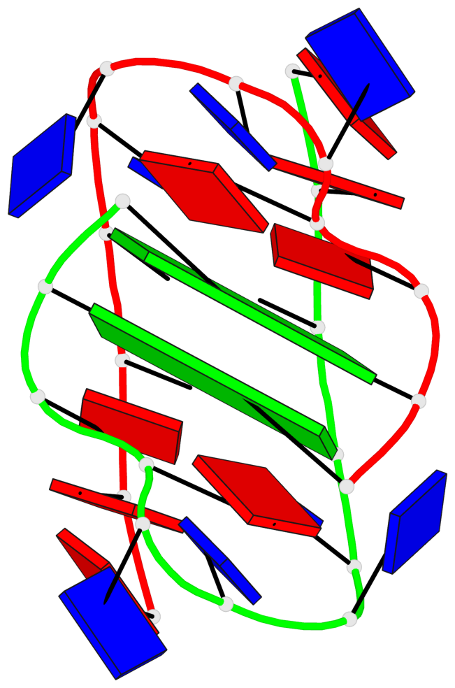
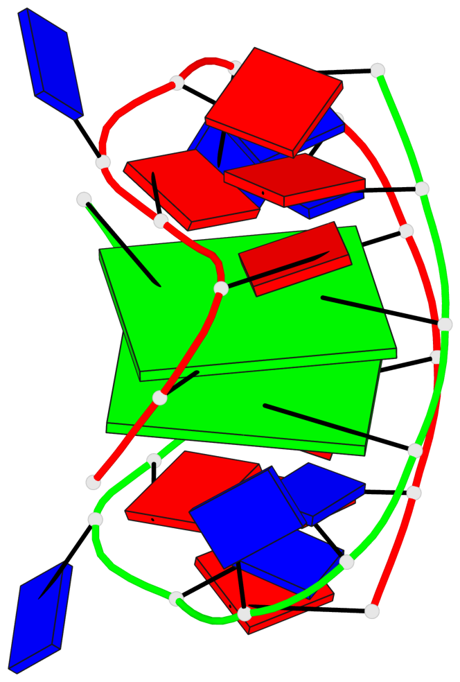
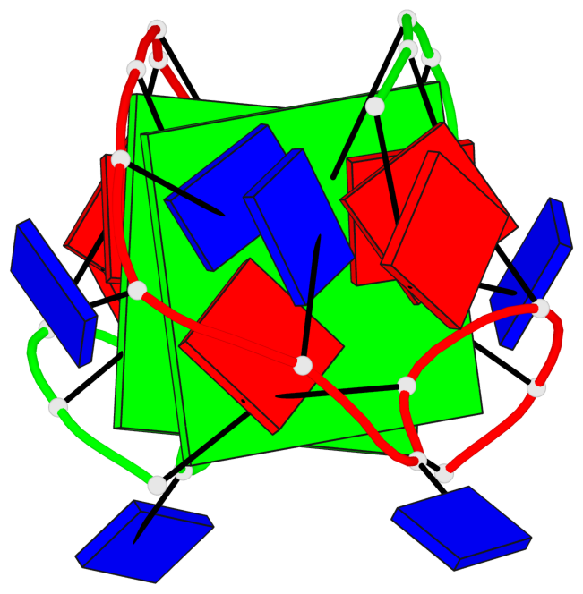
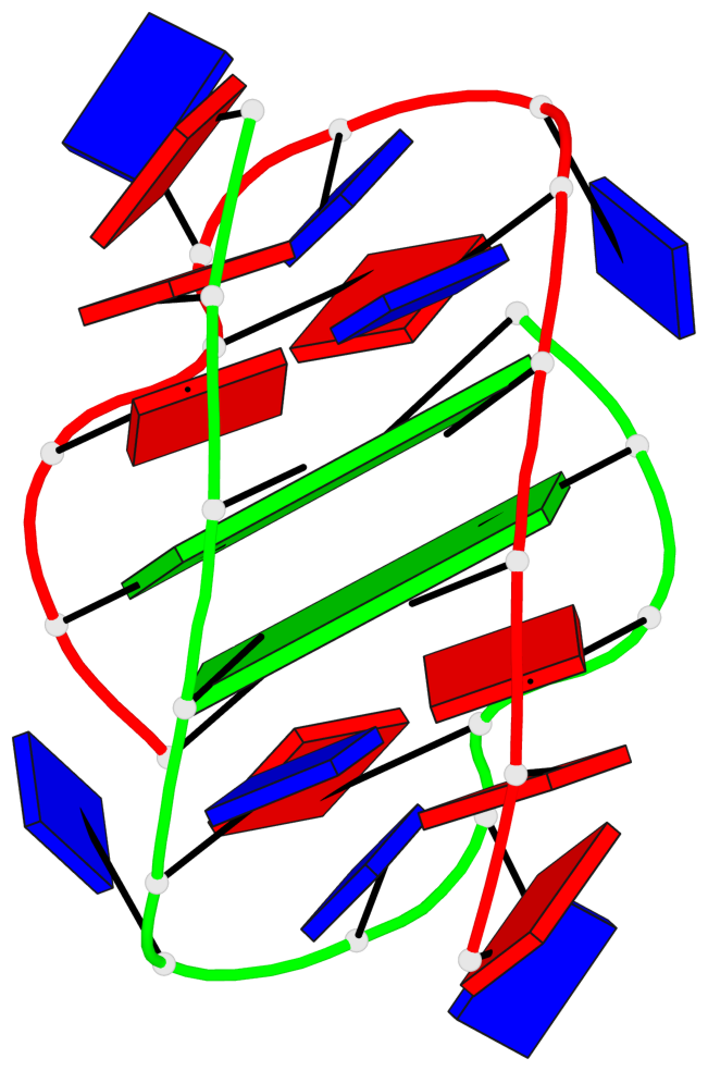
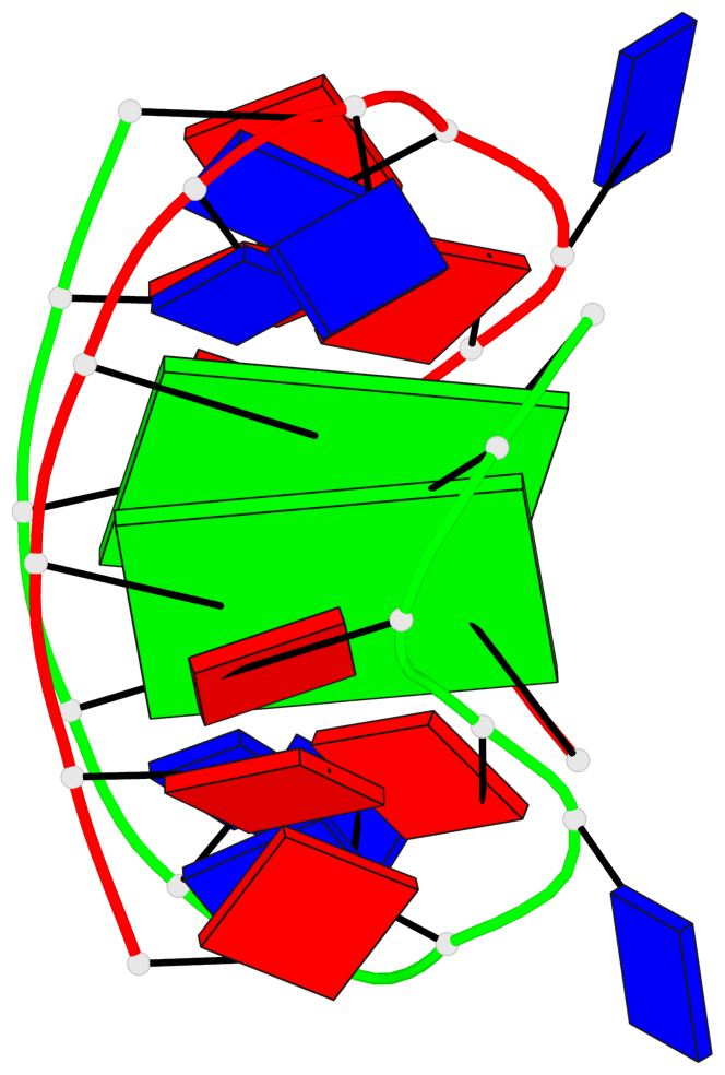
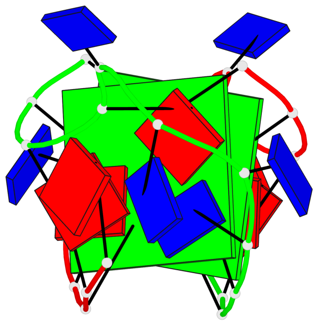
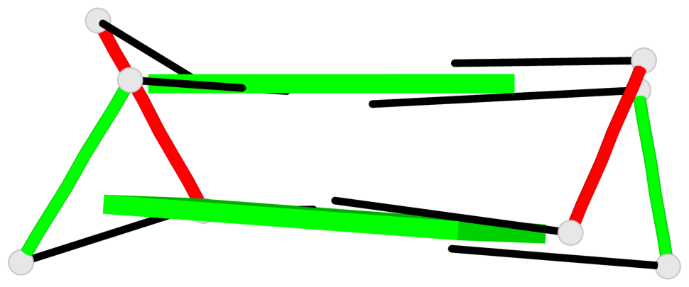
Base-block schematics in six views
List of 2 G-tetrads
 1 glyco-bond=ss-- sugar=-... groove=-w-n planarity=0.515 type=other  nts=4 GGGG A.DG3,B.DG11,A.DG12,B.DG4
 2 glyco-bond=--ss sugar=...- groove=-w-n planarity=0.516 type=other  nts=4 GGGG A.DG4,B.DG12,A.DG11,B.DG3
List of 1 G4-helix
In DSSR, a G4-helix is defined by stacking interactions of G-tetrads, regardless of backbone connectivity, and may contain more than one G4-stem.
Helix#1, 2 G-tetrad layers, inter-molecular, with 1 stem
 |
1 glyco-bond=ss-- sugar=-... groove=-w-n Major-->WC nts=4 GGGG A.DG3,B.DG11,A.DG12,B.DG4 2 glyco-bond=--ss sugar=...- groove=-w-n WC-->Major nts=4 GGGG A.DG4,B.DG12,A.DG11,B.DG3 step#1 mm(<>,outward) area=13.64 rise=3.56 twist=13.9 strand#1 DNA glyco-bond=s- sugar=-. nts=2 GG A.DG3,A.DG4 strand#2 DNA glyco-bond=s- sugar=.. nts=2 GG B.DG11,B.DG12 strand#3 DNA glyco-bond=-s sugar=.. nts=2 GG A.DG12,A.DG11 strand#4 DNA glyco-bond=-s sugar=.- nts=2 GG B.DG4,B.DG3 |
| 1 stacking diagram | |
 |
1 glyco-bond=ss-- sugar=-... groove=-w-n Major-->WC nts=4 GGGG A.DG3,B.DG11,A.DG12,B.DG4 |
List of 1 G4-stem
In DSSR, a G4-stem is defined as a G4-helix with backbone connectivity. Bulges are also allowed along each of the four strands.
Stem#1, 2 G-tetrad layers, 2 loops, inter-molecular, UUDD, anti-parallel, (2+2)
 |
1 glyco-bond=ss-- sugar=-... groove=-w-n Major-->WC nts=4 GGGG A.DG3,B.DG11,A.DG12,B.DG4 2 glyco-bond=--ss sugar=...- groove=-w-n WC-->Major nts=4 GGGG A.DG4,B.DG12,A.DG11,B.DG3 step#1 mm(<>,outward) area=13.64 rise=3.56 twist=13.9 strand#1 U DNA glyco-bond=s- sugar=-. nts=2 GG A.DG3,A.DG4 strand#2 U DNA glyco-bond=s- sugar=.. nts=2 GG B.DG11,B.DG12 strand#3 D DNA glyco-bond=-s sugar=.. nts=2 GG A.DG12,A.DG11 strand#4 D DNA glyco-bond=-s sugar=.- nts=2 GG B.DG4,B.DG3 loop#1 type=diagonal strands=[#1,#3] nts=6 TTTTAA A.DT5,A.DT6,A.DT7,A.DT8,A.DA9,A.DA10 loop#2 type=diagonal strands=[#4,#2] nts=6 TTTTAA B.DT5,B.DT6,B.DT7,B.DT8,B.DA9,B.DA10 |