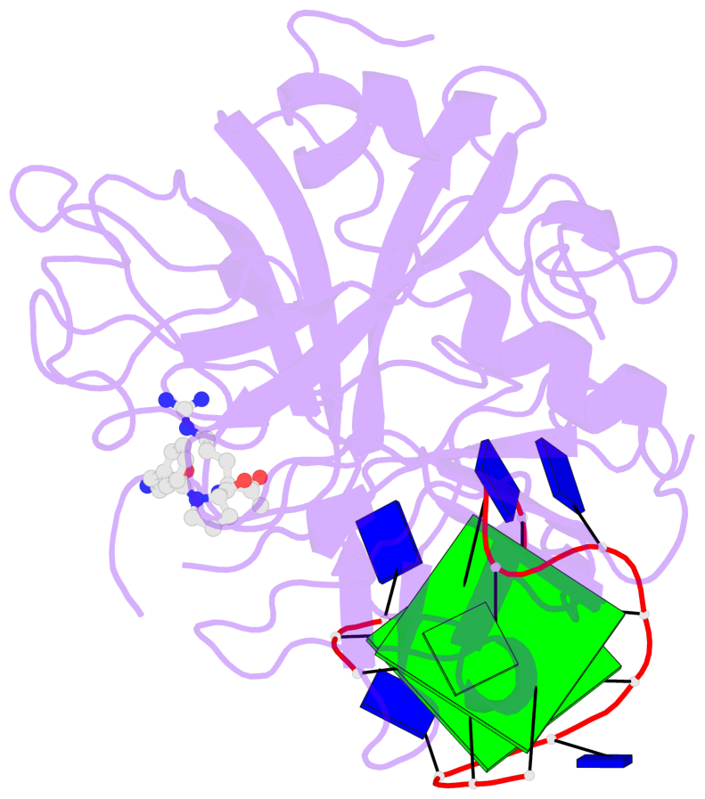
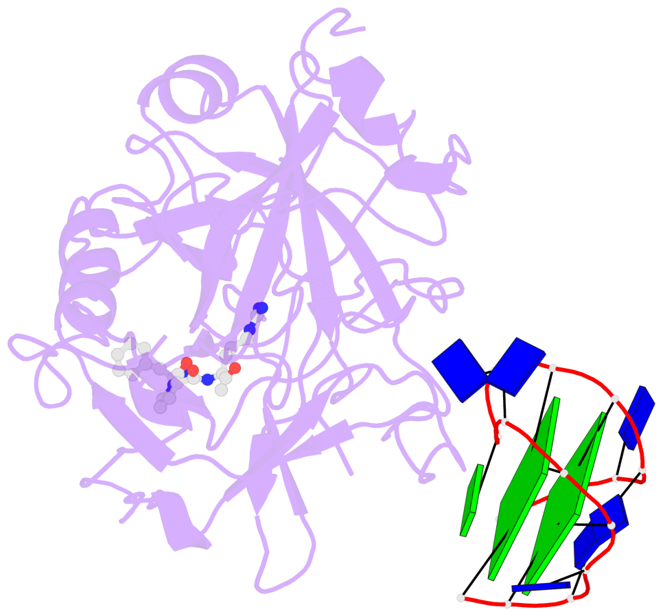
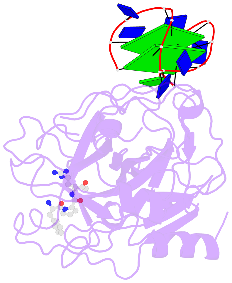
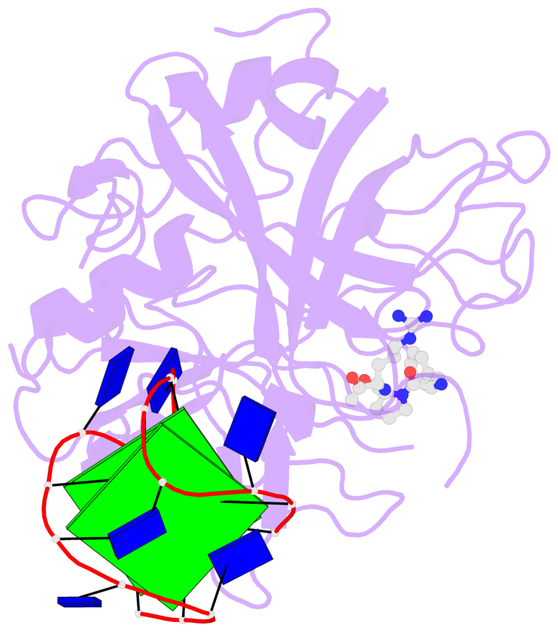
Detailed DSSR results for the G-quadruplex: PDB entry 1hut
Created and maintained by Xiang-Jun Lu <xiangjun@x3dna.org>
Citation: Please cite the NAR'20 DSSR-PyMOL schematics paper and/or the NAR'15 DSSR method paper.
Summary information
- PDB id
- 1hut
- Class
- hydrolase-hydrolase inhibitor-DNA
- Method
- X-ray (2.9 Å)
- Summary
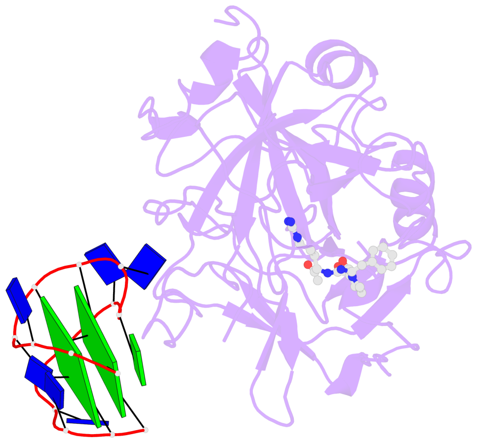
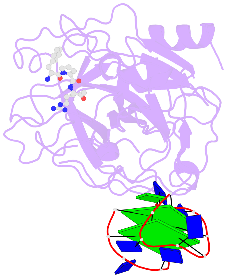
- The structure of alpha-thrombin inhibited by a 15-mer single-stranded DNA aptamer
- Reference
- Padmanabhan K, Padmanabhan KP, Ferrara JD, Sadler JE, Tulinsky A (1993): "The structure of alpha-thrombin inhibited by a 15-mer single-stranded DNA aptamer." J.Biol.Chem., 268, 17651-17654.
- Abstract
- The structure of a complex between human alpha-thrombin and a GGTTGGTGTGGTTGG 15-nucleotide consensus sequence has been solved by x-ray crystallography and refined at 2.9-A resolution to an R value of 0.159. As in solution, in the complex the single-stranded DNA folds into a structure with two G-quartets. The DNA is sandwiched between two different positively charged regions of two symmetry-related thrombin molecules in the crystal structure making ionic and hydrophobic interactions. One region is the fibrinogen recognition exosite and the other, the putative heparin binding site. The lack of inhibition of fibrinogen clotting and platelet activation by the DNA 15-mer with the Arg75-->Glu mutant of thrombin is consistent with the several salt bridges of the DNA in the fibrinogen exosite. The association of DNA with the heparin site of a neighboring molecule appears to simply compensate residual charge. Differences in the 15-mer loop conformations between the complex and NMR solution structures can be attributed to conformational changes upon thrombin binding. Although G-quadruplexes are favored in the presence of monovalent cations, there is no evidence of the latter in the thrombin complex.
- G4 notes
- 2 G-tetrads, 1 G4 helix, 1 G4 stem, 2(-Lw-Ln-Lw), chair(2+2), UDUD
Base-block schematics in six views
List of 2 G-tetrads
 1 glyco-bond=s-s- sugar=.-.3 groove=wnwn planarity=0.292 type=other  nts=4 GGGG D.DG401,D.DG406,D.DG410,D.DG415
 2 glyco-bond=-s-s sugar=.--. groove=wnwn planarity=0.452 type=other  nts=4 GGGG D.DG402,D.DG405,D.DG411,D.DG414
List of 1 G4-helix
In DSSR, a G4-helix is defined by stacking interactions of G-tetrads, regardless of backbone connectivity, and may contain more than one G4-stem.
Helix#1, 2 G-tetrad layers, INTRA-molecular, with 1 stem
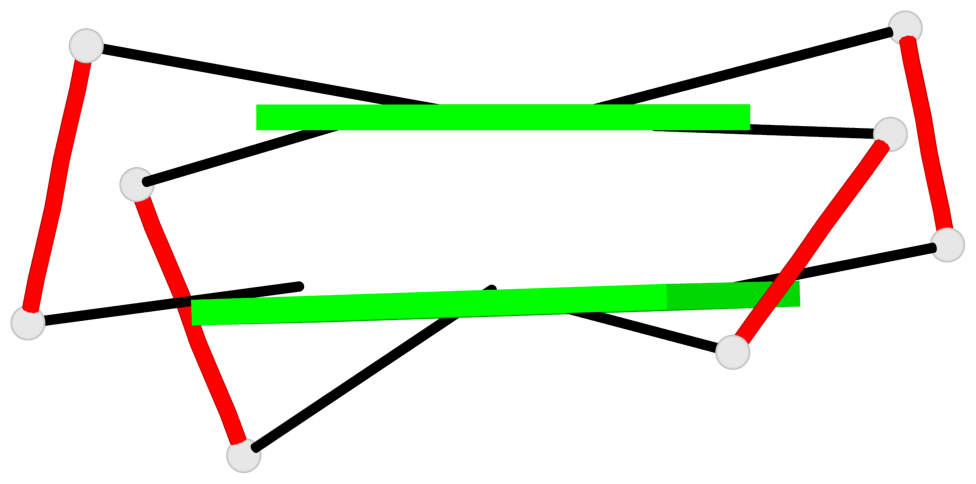 |
1 glyco-bond=s-s- sugar=.-.3 groove=wnwn Major-->WC nts=4 GGGG D.DG401,D.DG406,D.DG410,D.DG415 2 glyco-bond=-s-s sugar=.--. groove=wnwn WC-->Major nts=4 GGGG D.DG402,D.DG405,D.DG411,D.DG414 step#1 mm(<>,outward) area=14.48 rise=3.61 twist=15.6 strand#1 DNA glyco-bond=s- sugar=.. nts=2 GG D.DG401,D.DG402 strand#2 DNA glyco-bond=-s sugar=-- nts=2 GG D.DG406,D.DG405 strand#3 DNA glyco-bond=s- sugar=.- nts=2 GG D.DG410,D.DG411 strand#4 DNA glyco-bond=-s sugar=3. nts=2 GG D.DG415,D.DG414 |
| 1 stacking diagram | |
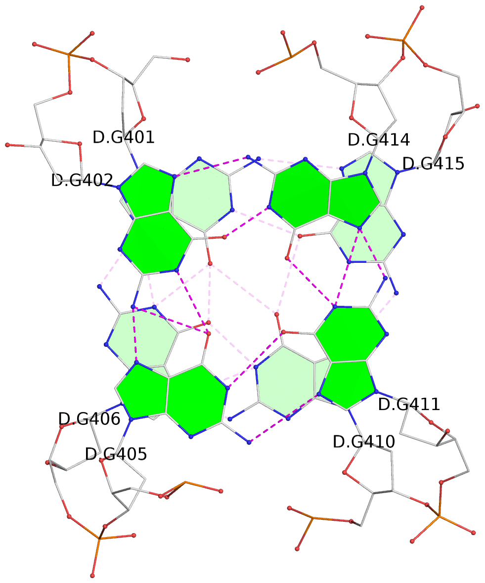 |
1 glyco-bond=s-s- sugar=.-.3 groove=wnwn Major-->WC nts=4 GGGG D.DG401,D.DG406,D.DG410,D.DG415 |
List of 1 G4-stem
In DSSR, a G4-stem is defined as a G4-helix with backbone connectivity. Bulges are also allowed along each of the four strands.
Stem#1, 2 G-tetrad layers, 3 loops, INTRA-molecular, UDUD, anti-parallel, 2(-Lw-Ln-Lw), chair(2+2)
 |
1 glyco-bond=s-s- sugar=.-.3 groove=wnwn Major-->WC nts=4 GGGG D.DG401,D.DG406,D.DG410,D.DG415 2 glyco-bond=-s-s sugar=.--. groove=wnwn WC-->Major nts=4 GGGG D.DG402,D.DG405,D.DG411,D.DG414 step#1 mm(<>,outward) area=14.48 rise=3.61 twist=15.6 strand#1 U DNA glyco-bond=s- sugar=.. nts=2 GG D.DG401,D.DG402 strand#2 D DNA glyco-bond=-s sugar=-- nts=2 GG D.DG406,D.DG405 strand#3 U DNA glyco-bond=s- sugar=.- nts=2 GG D.DG410,D.DG411 strand#4 D DNA glyco-bond=-s sugar=3. nts=2 GG D.DG415,D.DG414 loop#1 type=lateral strands=[#1,#2] nts=2 TT D.DT403,D.DT404 loop#2 type=lateral strands=[#2,#3] nts=3 TGT D.DT407,D.DG408,D.DT409 loop#3 type=lateral strands=[#3,#4] nts=2 TT D.DT412,D.DT413 |