Detailed DSSR results for the G-quadruplex: PDB entry 1j8g
Created and maintained by Xiang-Jun Lu <xiangjun@x3dna.org>
Citation: Please cite the NAR'20 DSSR-PyMOL schematics paper and/or the NAR'15 DSSR method paper.
Summary information
- PDB id
- 1j8g
- Class
- RNA
- Method
- X-ray (0.61 Å)
- Summary
- X-ray analysis of a RNA tetraplex r(uggggu)4 at ultra-high resolution
- Reference
- Deng J, Xiong Y, Sundaralingam M (2001): "X-ray analysis of an RNA tetraplex (UGGGGU)(4) with divalent Sr(2+) ions at subatomic resolution (0.61 A)." Proc.Natl.Acad.Sci.USA, 98, 13665-13670. doi: 10.1073/pnas.241374798.
- Abstract
- Four-stranded guanine tetraplexes in RNA have been identified to be involved in crucial biological functions, such as dimerization of retroviral RNA, translational repression, and mRNA turnover. However, the structural basis for these biological processes is still largely unknown. Here we report the RNA tetraplex structure (UGGGGU)(4) at ultra-high resolution (0.61 A). The space group is P42(1)2, and cell constants are a = b = 36.16 A and c = 74.09 A. The structure was solved by the multiple-wavelength anomalous dispersion method using a set of three-wavelength data of the isomorphous bromo derivative (br)UGGGGU and refined to 0.61-A resolution. Each of the four strands in the asymmetric unit forms a parallel tetraplex with symmetry-related molecules. The tetraplex molecules stack on one another in opposite polarity (head-to-head or tail-to-tail) to form a pseudocontinuous column. All of the 5'-end uridines rotate around the backbone of G2, swing out, and form unique octaplexes with the neighboring G tetraplexes, whereas the 3'-end uridines are stacked-in and form uridine tetrads. All of the bases are anti, and the riboses are in the mixed C2'- and C3'-puckering mode. Strontium ions are observed in every other guanine tetrad plane, sitting on the fourfold axis and associated to the eight O6 atoms of neighboring guanine bases in a bipyramidal-antiprism geometry. The hydrogens are clearly observed in the structure.
- G4 notes
- 4 G-tetrads, 1 G4 helix, 1 G4 stem, parallel(4+0), UUUU
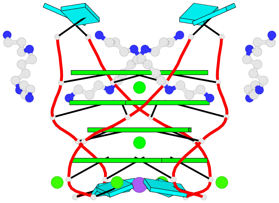
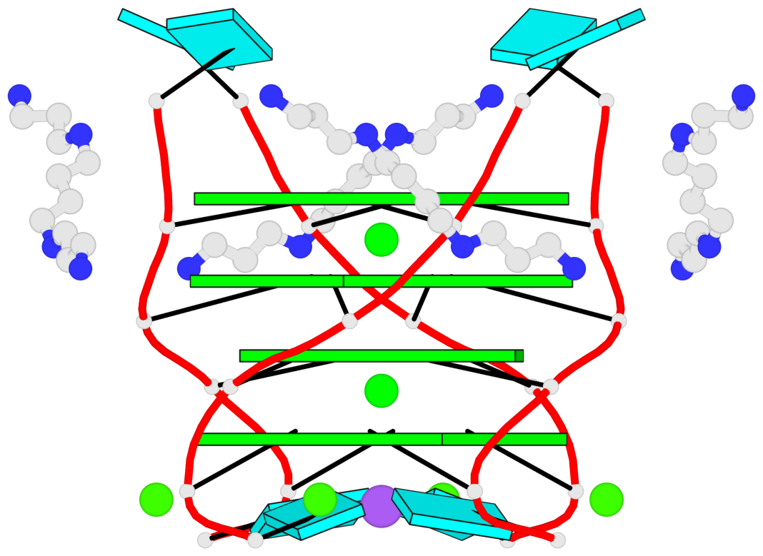
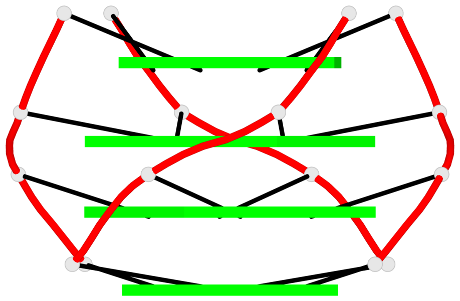
Base-block schematics in six views
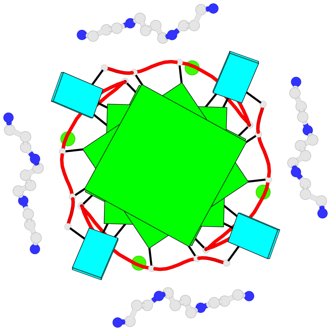


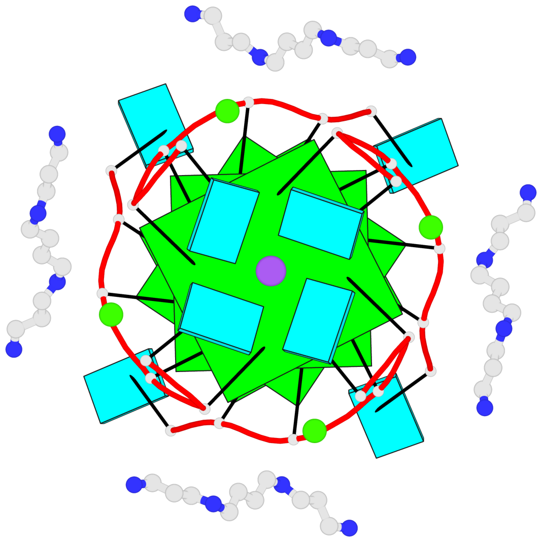
List of 4 G-tetrads
 1 glyco-bond=---- sugar=3333 groove=---- planarity=0.238 type=other  nts=4 GGGG 1:A.G2,3:A.G2,2:A.G2,4:A.G2
 2 glyco-bond=---- sugar=---- groove=---- planarity=0.246 type=bowl   nts=4 GGGG 1:A.G3,3:A.G3,2:A.G3,4:A.G3
 3 glyco-bond=---- sugar=3333 groove=---- planarity=0.123 type=planar nts=4 GGGG 1:A.G4,3:A.G4,2:A.G4,4:A.G4
 4 glyco-bond=---- sugar=3333 groove=---- planarity=0.295 type=bowl   nts=4 GGGG 1:A.G5,3:A.G5,2:A.G5,4:A.G5
List of 1 G4-helix
In DSSR, a G4-helix is defined by stacking interactions of G-tetrads, regardless of backbone connectivity, and may contain more than one G4-stem.
Helix#1, 4 G-tetrad layers, inter-molecular, with 1 stem
 |
1 glyco-bond=---- sugar=3333 groove=---- WC-->Major nts=4 GGGG 1:A.G2,3:A.G2,2:A.G2,4:A.G2 2 glyco-bond=---- sugar=---- groove=---- WC-->Major nts=4 GGGG 1:A.G3,3:A.G3,2:A.G3,4:A.G3 3 glyco-bond=---- sugar=3333 groove=---- WC-->Major nts=4 GGGG 1:A.G4,3:A.G4,2:A.G4,4:A.G4 4 glyco-bond=---- sugar=3333 groove=---- WC-->Major nts=4 GGGG 1:A.G5,3:A.G5,2:A.G5,4:A.G5 step#1 pm(>>,forward) area=11.74 rise=3.47 twist=27.5 step#2 pm(>>,forward) area=8.71 rise=3.15 twist=35.4 step#3 pm(>>,forward) area=12.54 rise=3.52 twist=25.2 strand#1 RNA glyco-bond=---- sugar=3-33 nts=4 GGGG 1:A.G2,1:A.G3,1:A.G4,1:A.G5 strand#2 RNA glyco-bond=---- sugar=3-33 nts=4 GGGG 3:A.G2,3:A.G3,3:A.G4,3:A.G5 strand#3 RNA glyco-bond=---- sugar=3-33 nts=4 GGGG 2:A.G2,2:A.G3,2:A.G4,2:A.G5 strand#4 RNA glyco-bond=---- sugar=3-33 nts=4 GGGG 4:A.G2,4:A.G3,4:A.G4,4:A.G5 |
| 3 stacking diagrams | |
 |
1 glyco-bond=---- sugar=3333 groove=---- WC-->Major nts=4 GGGG 1:A.G2,3:A.G2,2:A.G2,4:A.G2 |
 |
2 glyco-bond=---- sugar=---- groove=---- WC-->Major nts=4 GGGG 1:A.G3,3:A.G3,2:A.G3,4:A.G3 |
 |
3 glyco-bond=---- sugar=3333 groove=---- WC-->Major nts=4 GGGG 1:A.G4,3:A.G4,2:A.G4,4:A.G4 |
List of 1 G4-stem
In DSSR, a G4-stem is defined as a G4-helix with backbone connectivity. Bulges are also allowed along each of the four strands.
Stem#1, 4 G-tetrad layers, 0 loops, inter-molecular, UUUU, parallel, parallel(4+0)
 |
1 glyco-bond=---- sugar=3333 groove=---- WC-->Major nts=4 GGGG 1:A.G2,3:A.G2,2:A.G2,4:A.G2 2 glyco-bond=---- sugar=---- groove=---- WC-->Major nts=4 GGGG 1:A.G3,3:A.G3,2:A.G3,4:A.G3 3 glyco-bond=---- sugar=3333 groove=---- WC-->Major nts=4 GGGG 1:A.G4,3:A.G4,2:A.G4,4:A.G4 4 glyco-bond=---- sugar=3333 groove=---- WC-->Major nts=4 GGGG 1:A.G5,3:A.G5,2:A.G5,4:A.G5 step#1 pm(>>,forward) area=11.74 rise=3.47 twist=27.5 step#2 pm(>>,forward) area=8.71 rise=3.15 twist=35.4 step#3 pm(>>,forward) area=12.54 rise=3.52 twist=25.2 strand#1 U RNA glyco-bond=---- sugar=3-33 nts=4 GGGG 1:A.G2,1:A.G3,1:A.G4,1:A.G5 strand#2 U RNA glyco-bond=---- sugar=3-33 nts=4 GGGG 3:A.G2,3:A.G3,3:A.G4,3:A.G5 strand#3 U RNA glyco-bond=---- sugar=3-33 nts=4 GGGG 2:A.G2,2:A.G3,2:A.G4,2:A.G5 strand#4 U RNA glyco-bond=---- sugar=3-33 nts=4 GGGG 4:A.G2,4:A.G3,4:A.G4,4:A.G5 |