Detailed DSSR results for the G-quadruplex: PDB entry 1jpq
Created and maintained by Xiang-Jun Lu <xiangjun@x3dna.org>
Citation: Please cite the NAR'20 DSSR-PyMOL schematics paper and/or the NAR'15 DSSR method paper.
Summary information
- PDB id
- 1jpq
- Class
- DNA
- Method
- X-ray (1.6 Å)
- Summary
- Crystal structure of the oxytricha telomeric DNA at 1.6a
- Reference
- Haider S, Parkinson GN, Neidle S (2002): "Crystal structure of the potassium form of an Oxytricha nova G-quadruplex." J.Mol.Biol., 320, 189-200. doi: 10.1016/S0022-2836(02)00428-X.
- Abstract
- The crystal structures of the potassium-containing quadruplex formed from the Oxytricha nova sequence d(GGGGTTTTGGGG) are reported, in two space groups, the orthorhombic P2(1)2(1)2(1) and the trigonal P3(2)21, which diffract to 2.0 A and 1.49 A, respectively. The orthorhombic form contains two independent quadruplexes in the asymmetric unit, and the trigonal form contains one. All three of these quadruplexes adopt an identical fold, with two strands forming an antiparallel diagonal arrangement. This is identical with that observed previously in NMR studies of the native sodium and potassium forms, and a crystallographic analysis of it complexed with an O. nova protein. The present analysis demonstrates that the native structure is the same in solution and in the crystalline state and, moreover, that the nature of the counter-ion does not affect the overall fold of this quadruplex. The analysis corrects an earlier crystallographic study of this quadruplex. The conformation of the tetra-thymine loop is described in detail, which involves the third thymine base folding back to interact with the first thymine base. The water networks in the grooves and loops are described and, in particular, the ability of water molecules to form a continuous spine of hydration in the narrow groove is detailed. Each quadruplex has five potassium ions organised in a linear channel, with square antiprismatic coordination to each ion from oxygen atoms.
- G4 notes
- 4 G-tetrads, 1 G4 helix, 1 G4 stem, (2+2), UDDU
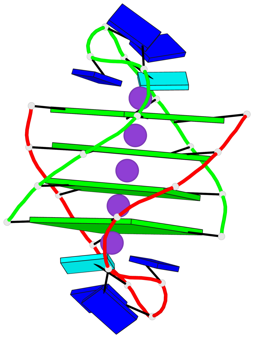
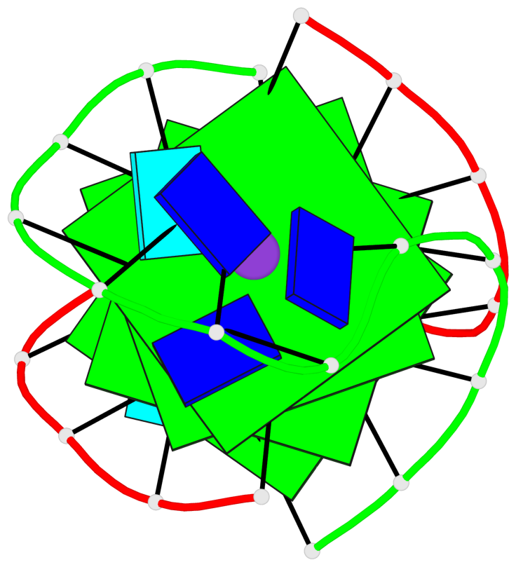
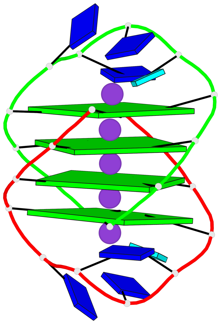
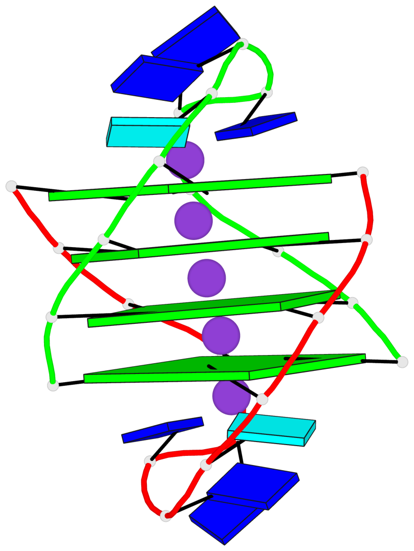
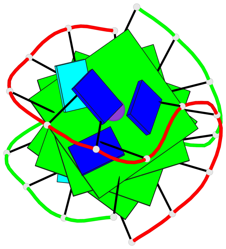
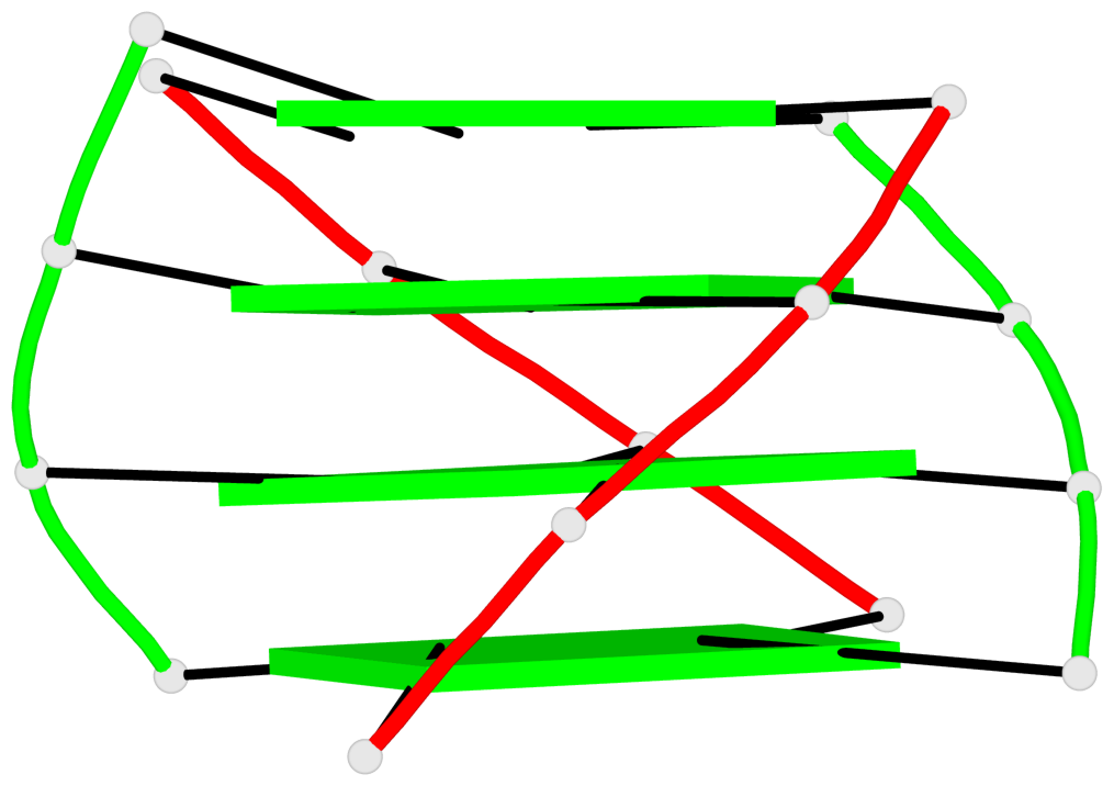
Base-block schematics in six views

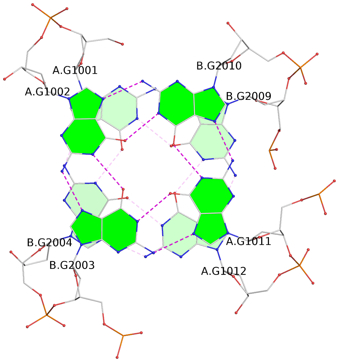
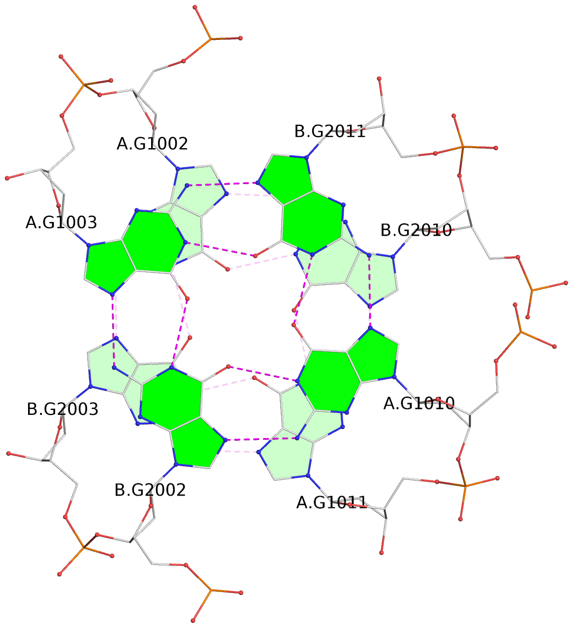
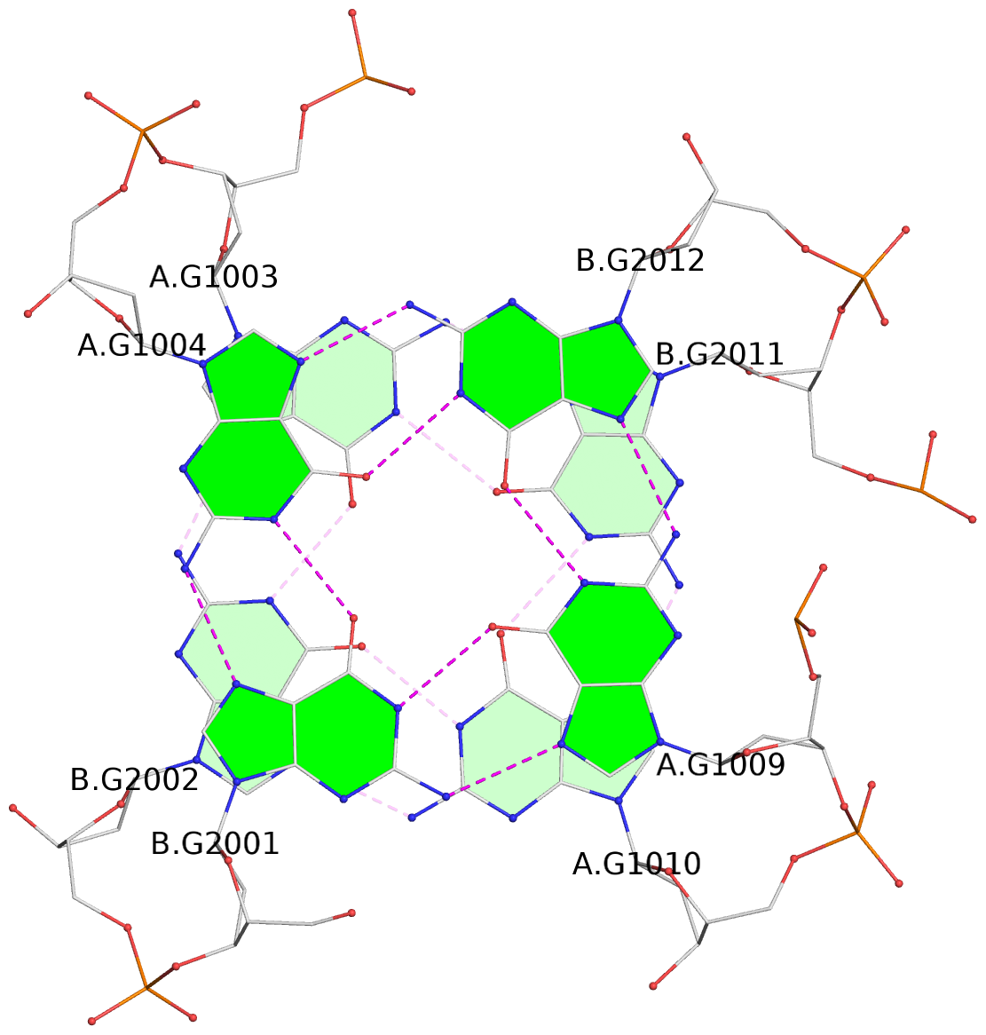
List of 4 G-tetrads
 1 glyco-bond=s--s sugar=---- groove=w-n- planarity=0.283 type=bowl   nts=4 GGGG A.DG1001,B.DG2004,A.DG1012,B.DG2009
 2 glyco-bond=-ss- sugar=---- groove=w-n- planarity=0.165 type=other  nts=4 GGGG A.DG1002,B.DG2003,A.DG1011,B.DG2010
 3 glyco-bond=s--s sugar=---- groove=w-n- planarity=0.154 type=planar nts=4 GGGG A.DG1003,B.DG2002,A.DG1010,B.DG2011
 4 glyco-bond=-ss- sugar=---- groove=w-n- planarity=0.239 type=other  nts=4 GGGG A.DG1004,B.DG2001,A.DG1009,B.DG2012
List of 1 G4-helix
In DSSR, a G4-helix is defined by stacking interactions of G-tetrads, regardless of backbone connectivity, and may contain more than one G4-stem.
Helix#1, 4 G-tetrad layers, inter-molecular, with 1 stem
 |
1 glyco-bond=s--s sugar=---- groove=w-n- Major-->WC nts=4 GGGG A.DG1001,B.DG2004,A.DG1012,B.DG2009 2 glyco-bond=-ss- sugar=---- groove=w-n- WC-->Major nts=4 GGGG A.DG1002,B.DG2003,A.DG1011,B.DG2010 3 glyco-bond=s--s sugar=---- groove=w-n- Major-->WC nts=4 GGGG A.DG1003,B.DG2002,A.DG1010,B.DG2011 4 glyco-bond=-ss- sugar=---- groove=w-n- WC-->Major nts=4 GGGG A.DG1004,B.DG2001,A.DG1009,B.DG2012 step#1 mm(<>,outward) area=14.57 rise=3.50 twist=16.9 step#2 pp(><,inward) area=19.29 rise=3.53 twist=36.6 step#3 mm(<>,outward) area=12.90 rise=3.47 twist=18.2 strand#1 DNA glyco-bond=s-s- sugar=---- nts=4 GGGG A.DG1001,A.DG1002,A.DG1003,A.DG1004 strand#2 DNA glyco-bond=-s-s sugar=---- nts=4 GGGG B.DG2004,B.DG2003,B.DG2002,B.DG2001 strand#3 DNA glyco-bond=-s-s sugar=---- nts=4 GGGG A.DG1012,A.DG1011,A.DG1010,A.DG1009 strand#4 DNA glyco-bond=s-s- sugar=---- nts=4 GGGG B.DG2009,B.DG2010,B.DG2011,B.DG2012 |
| 3 stacking diagrams | |
 |
1 glyco-bond=s--s sugar=---- groove=w-n- Major-->WC nts=4 GGGG A.DG1001,B.DG2004,A.DG1012,B.DG2009 |
 |
2 glyco-bond=-ss- sugar=---- groove=w-n- WC-->Major nts=4 GGGG A.DG1002,B.DG2003,A.DG1011,B.DG2010 |
 |
3 glyco-bond=s--s sugar=---- groove=w-n- Major-->WC nts=4 GGGG A.DG1003,B.DG2002,A.DG1010,B.DG2011 |
List of 1 G4-stem
In DSSR, a G4-stem is defined as a G4-helix with backbone connectivity. Bulges are also allowed along each of the four strands.
Stem#1, 4 G-tetrad layers, 2 loops, inter-molecular, UDDU, anti-parallel, (2+2)
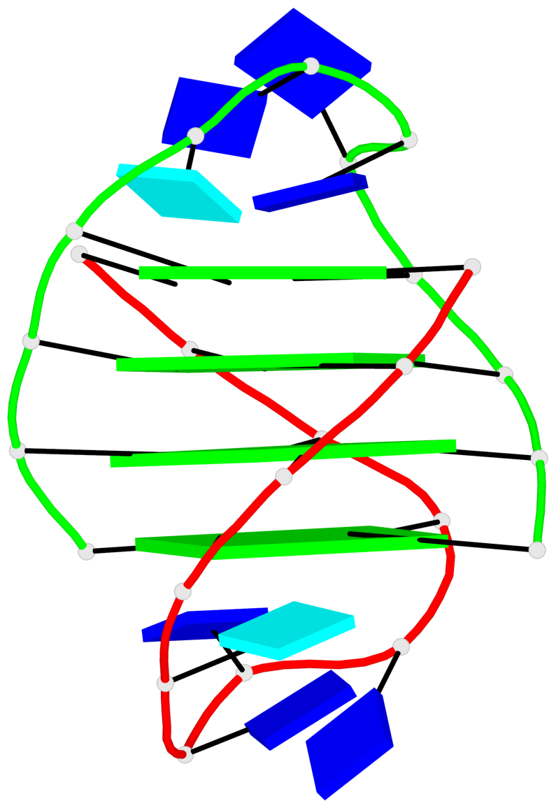 |
1 glyco-bond=s--s sugar=---- groove=w-n- Major-->WC nts=4 GGGG A.DG1001,B.DG2004,A.DG1012,B.DG2009 2 glyco-bond=-ss- sugar=---- groove=w-n- WC-->Major nts=4 GGGG A.DG1002,B.DG2003,A.DG1011,B.DG2010 3 glyco-bond=s--s sugar=---- groove=w-n- Major-->WC nts=4 GGGG A.DG1003,B.DG2002,A.DG1010,B.DG2011 4 glyco-bond=-ss- sugar=---- groove=w-n- WC-->Major nts=4 GGGG A.DG1004,B.DG2001,A.DG1009,B.DG2012 step#1 mm(<>,outward) area=14.57 rise=3.50 twist=16.9 step#2 pp(><,inward) area=19.29 rise=3.53 twist=36.6 step#3 mm(<>,outward) area=12.90 rise=3.47 twist=18.2 strand#1 U DNA glyco-bond=s-s- sugar=---- nts=4 GGGG A.DG1001,A.DG1002,A.DG1003,A.DG1004 strand#2 D DNA glyco-bond=-s-s sugar=---- nts=4 GGGG B.DG2004,B.DG2003,B.DG2002,B.DG2001 strand#3 D DNA glyco-bond=-s-s sugar=---- nts=4 GGGG A.DG1012,A.DG1011,A.DG1010,A.DG1009 strand#4 U DNA glyco-bond=s-s- sugar=---- nts=4 GGGG B.DG2009,B.DG2010,B.DG2011,B.DG2012 loop#1 type=diagonal strands=[#1,#3] nts=4 uTTT A.BRU1005,A.DT1006,A.DT1007,A.DT1008 loop#2 type=diagonal strands=[#2,#4] nts=4 uTTT B.BRU2005,B.DT2006,B.DT2007,B.DT2008 |