Detailed DSSR results for the G-quadruplex: PDB entry 1rde
Created and maintained by Xiang-Jun Lu <xiangjun@x3dna.org>
Citation: Please cite the NAR'20 DSSR-PyMOL schematics paper and/or the NAR'15 DSSR method paper.
Summary information
- PDB id
- 1rde
- Class
- DNA
- Method
- NMR
- Summary
- NMR structure of the thrombin-binding DNA aptamer stabilized by sr2+
- Reference
- Mao X, Marky LA, Gmeiner WH (2004): "NMR structure of the thrombin-binding DNA aptamer stabilized by Sr2+." J.Biomol.Struct.Dyn., 22, 25-33.
- Abstract
- The structure of thrombin-binding DNA aptamer complexed with a single Sr2+ ion (Sr2+:TBA complex) has been determined using NMR spectroscopy and restrained molecular dynamics simulations. The quadruplex structure for the Sr2+:TBA complex is similar in topology, but distinct in structure, from that previously reported for the K+:TBA complex. The inter-tetrad distance of the Sr2+:TBA complex is 3.8 angstroms, or 0.7 angstroms larger than in the K+:TBA complex. This substantial difference can be attributed to a different binding site for Sr2+ in the Sr2+:TBA complex than for K+ in the K+:TBA complex. The Sr2+:TBA complex assumes a 1:1 stoichiometry, and it is very likely that the Sr2+ ion simultaneously interacts with the eight O6 atoms of the two G-tetrads. The results indicate that quadruplex DNA structures are highly sensitive to the presence of specific metal ions. The binding of specific metal ions may modulate the biological activity of quadruplex DNA structures in vivo.
- G4 notes
- 2 G-tetrads, 1 G4 helix, 1 G4 stem, 2(+Ln+Lw+Ln), chair(2+2), UDUD
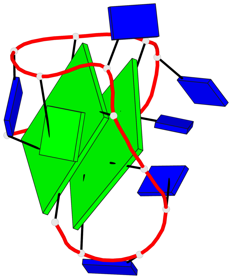
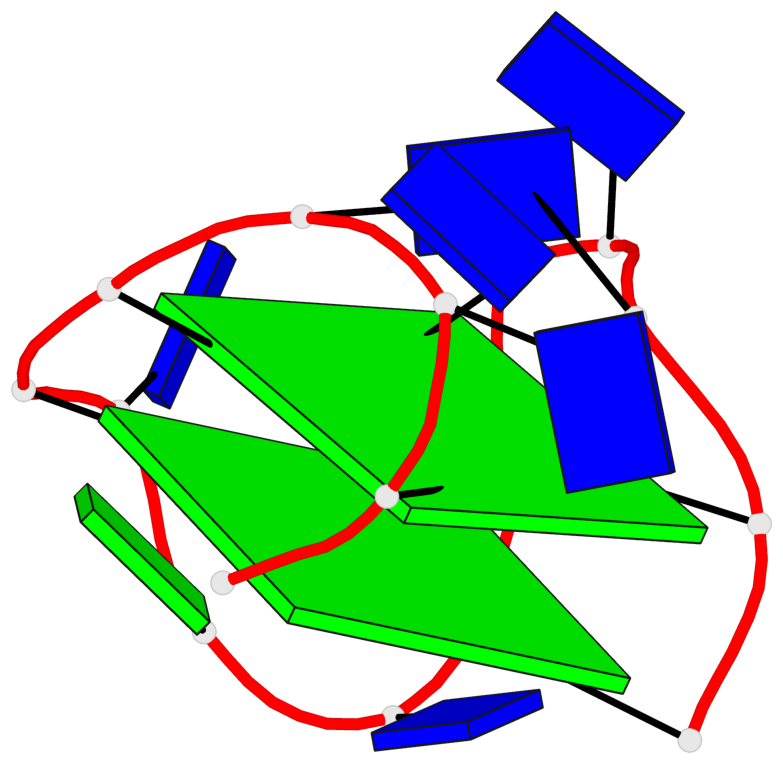
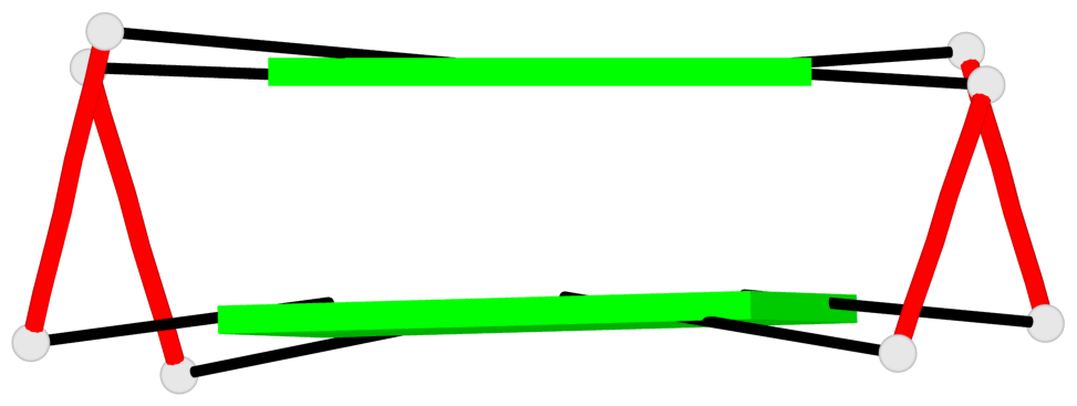
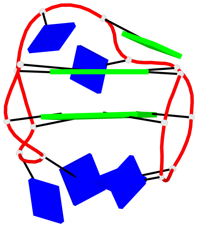
Base-block schematics in six views
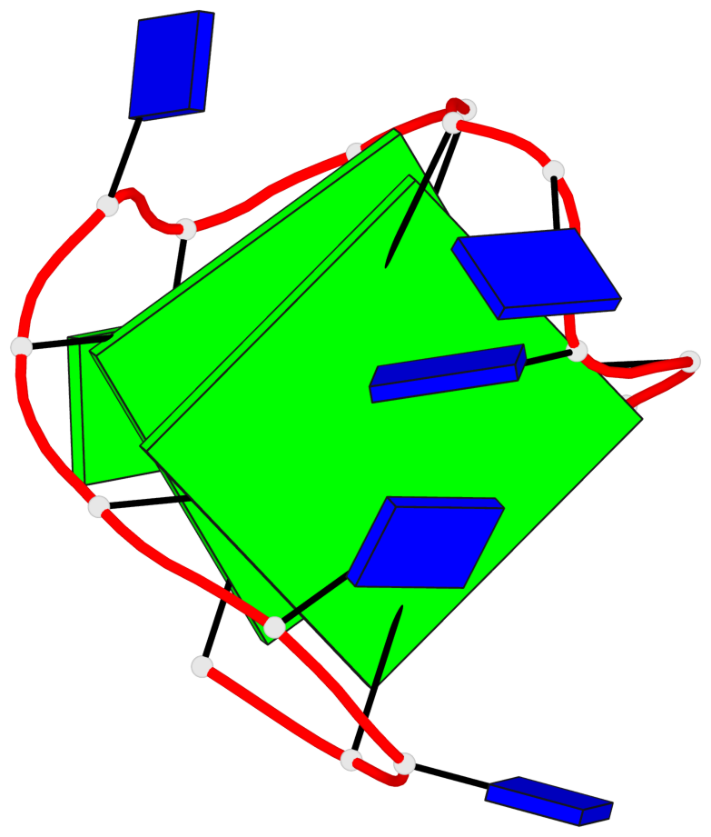
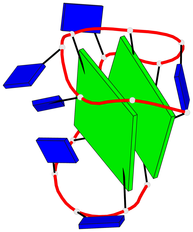
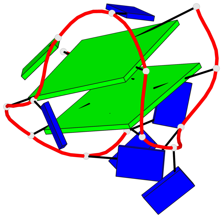
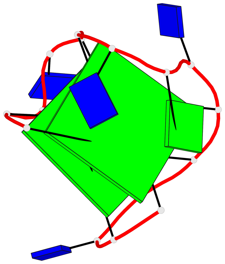
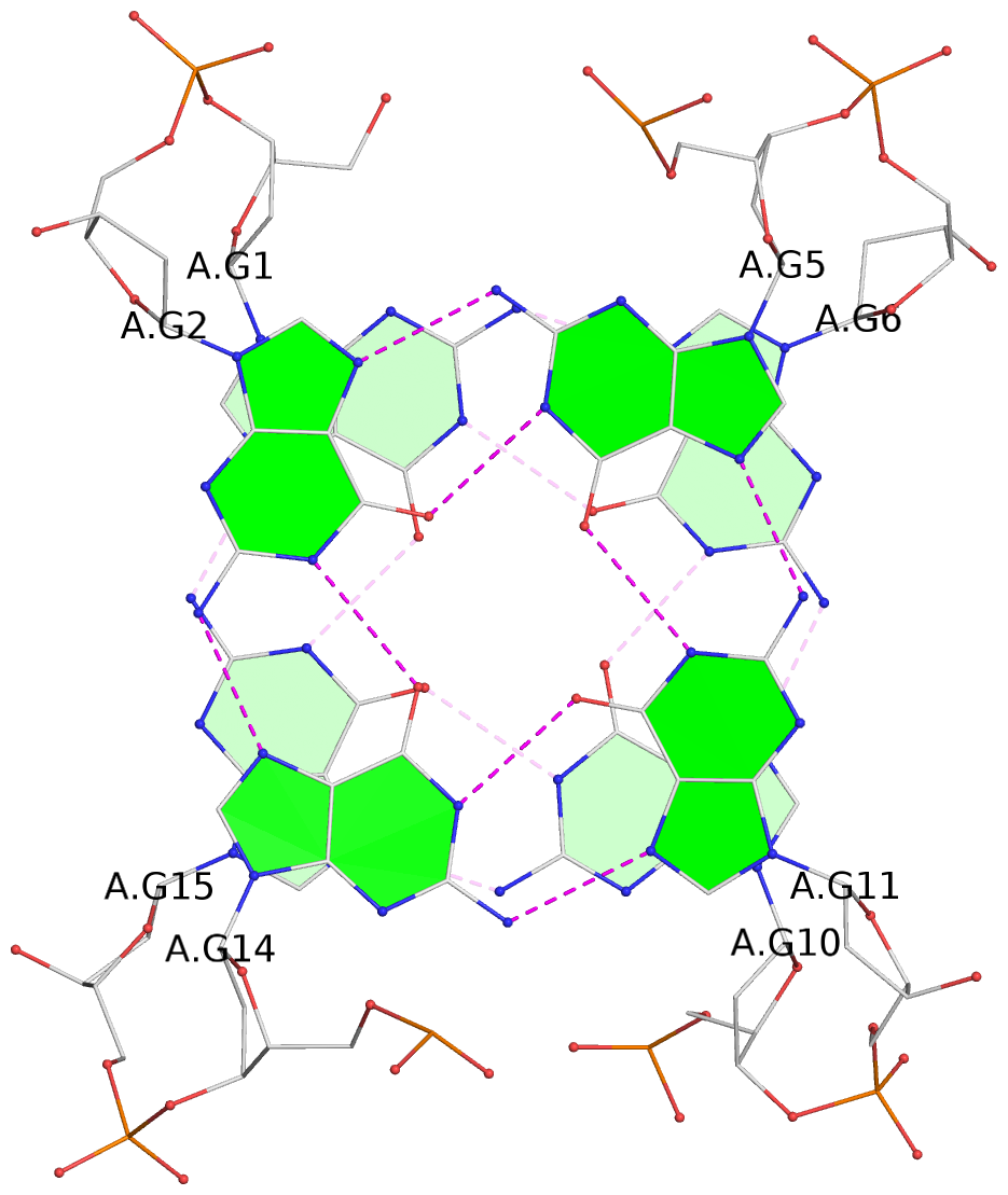
List of 2 G-tetrads
 1 glyco-bond=s-s- sugar=---- groove=wnwn planarity=0.178 type=other  nts=4 GGGG A.DG1,A.DG15,A.DG10,A.DG6
 2 glyco-bond=-s-s sugar=---- groove=wnwn planarity=0.254 type=bowl   nts=4 GGGG A.DG2,A.DG14,A.DG11,A.DG5
List of 1 G4-helix
In DSSR, a G4-helix is defined by stacking interactions of G-tetrads, regardless of backbone connectivity, and may contain more than one G4-stem.
Helix#1, 2 G-tetrad layers, INTRA-molecular, with 1 stem
 |
1 glyco-bond=s-s- sugar=---- groove=wnwn Major-->WC nts=4 GGGG A.DG1,A.DG15,A.DG10,A.DG6 2 glyco-bond=-s-s sugar=---- groove=wnwn WC-->Major nts=4 GGGG A.DG2,A.DG14,A.DG11,A.DG5 step#1 mm(<>,outward) area=17.53 rise=4.29 twist=11.3 strand#1 DNA glyco-bond=s- sugar=-- nts=2 GG A.DG1,A.DG2 strand#2 DNA glyco-bond=-s sugar=-- nts=2 GG A.DG15,A.DG14 strand#3 DNA glyco-bond=s- sugar=-- nts=2 GG A.DG10,A.DG11 strand#4 DNA glyco-bond=-s sugar=-- nts=2 GG A.DG6,A.DG5 |
| 1 stacking diagram | |
 |
1 glyco-bond=s-s- sugar=---- groove=wnwn Major-->WC nts=4 GGGG A.DG1,A.DG15,A.DG10,A.DG6 |
List of 1 G4-stem
In DSSR, a G4-stem is defined as a G4-helix with backbone connectivity. Bulges are also allowed along each of the four strands.
Stem#1, 2 G-tetrad layers, 3 loops, INTRA-molecular, UDUD, anti-parallel, 2(+Ln+Lw+Ln), chair(2+2)
 |
1 glyco-bond=s-s- sugar=---- groove=wnwn Major-->WC nts=4 GGGG A.DG1,A.DG15,A.DG10,A.DG6 2 glyco-bond=-s-s sugar=---- groove=wnwn WC-->Major nts=4 GGGG A.DG2,A.DG14,A.DG11,A.DG5 step#1 mm(<>,outward) area=17.53 rise=4.29 twist=11.3 strand#1 U DNA glyco-bond=s- sugar=-- nts=2 GG A.DG1,A.DG2 strand#2 D DNA glyco-bond=-s sugar=-- nts=2 GG A.DG15,A.DG14 strand#3 U DNA glyco-bond=s- sugar=-- nts=2 GG A.DG10,A.DG11 strand#4 D DNA glyco-bond=-s sugar=-- nts=2 GG A.DG6,A.DG5 loop#1 type=lateral strands=[#1,#4] nts=2 TT A.DT3,A.DT4 loop#2 type=lateral strands=[#4,#3] nts=3 TGT A.DT7,A.DG8,A.DT9 loop#3 type=lateral strands=[#3,#2] nts=2 TT A.DT12,A.DT13 |