Detailed DSSR results for the G-quadruplex: PDB entry 2avh
Created and maintained by Xiang-Jun Lu <xiangjun@x3dna.org>
Citation: Please cite the NAR'20 DSSR-PyMOL schematics paper and/or the NAR'15 DSSR method paper.
Summary information
- PDB id
- 2avh
- Class
- DNA
- Method
- X-ray (1.5 Å)
- Summary
- G4t3g4 dimeric quadruplex structure
- Reference
- Hazel P, Parkinson GN, Neidle S (2006): "Topology variation and loop structural homology in crystal and simulated structures of a bimolecular DNA quadruplex." J.Am.Chem.Soc., 128, 5480-5487. doi: 10.1021/ja058577+.
- Abstract
- The topology of DNA quadruplexes depends on the nature and number of the nucleotides linking G-quartet motifs. To assess the effects of a three-nucleotide TTT linker, the crystal structure of the DNA sequence d(G(4)T(3)G(4)) has been determined at 1.5 A resolution, together with that of the brominated analogue d(G(4)(Br)UTTG(4)) at 2.4 A resolution. Both sequences form bimolecular intermolecular G-quadruplexes with lateral loops. d(G(4)(Br)UTTG(4)) crystallized in the monoclinic space group P2(1) with three quadruplex molecules in the asymmetric unit, two associating together as a head-to-head stacked dimer, and the third as a single head-to-tail dimer. The head-to-head dimers have two lateral loops on the same G-quadruplex face and form an eight-G-quartet stack, with a linear array of seven K(+) ions between the quartets. d(G(4)T(3)G(4)) crystallized in the orthorhombic space group C222 and has a structure very similar to the head-to-tail dimer in the P2(1) unit cell. The sequence studied here is able to form several different folds; however, all four quadruplexes in the two structures have lateral loops, in contrast to the diagonal loops reported for the analogous quadruplex with T(4) loops. A total of seven independent T(3) loops were observed in the two structures. These can be classified into two discrete conformational classes, suggesting that these represent preferred loop conformations that are independent of crystal-packing forces.
- G4 notes
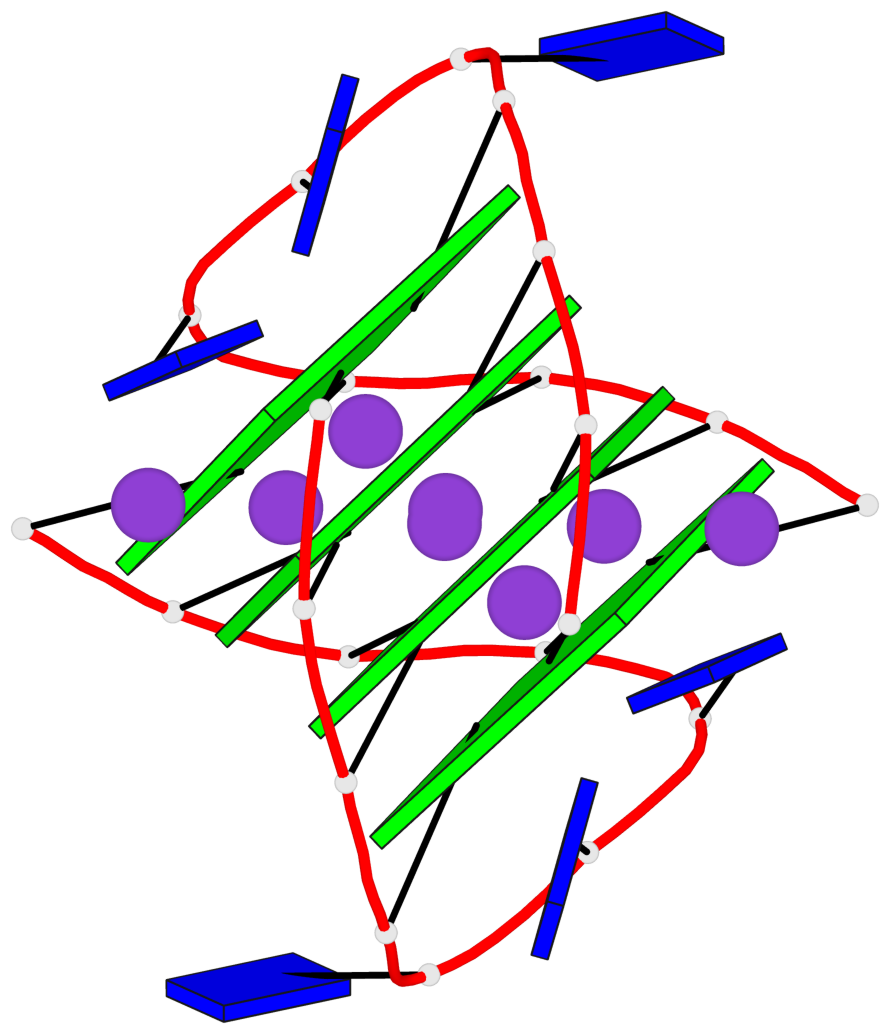
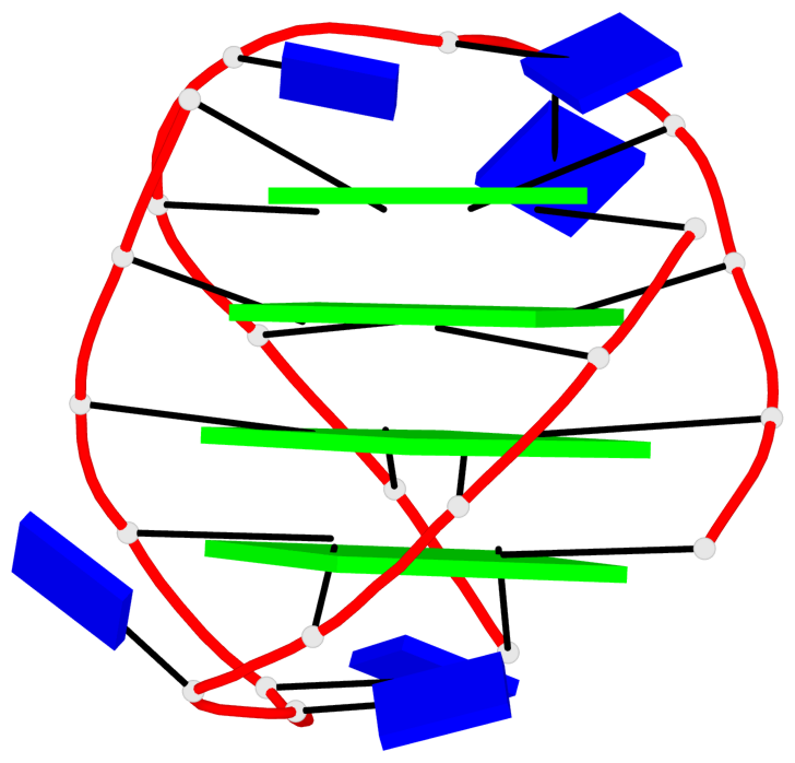
- 4 G-tetrads, 1 G4 helix, 1 G4 stem, (2+2), UDUD
Base-block schematics in six views
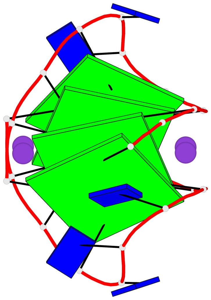
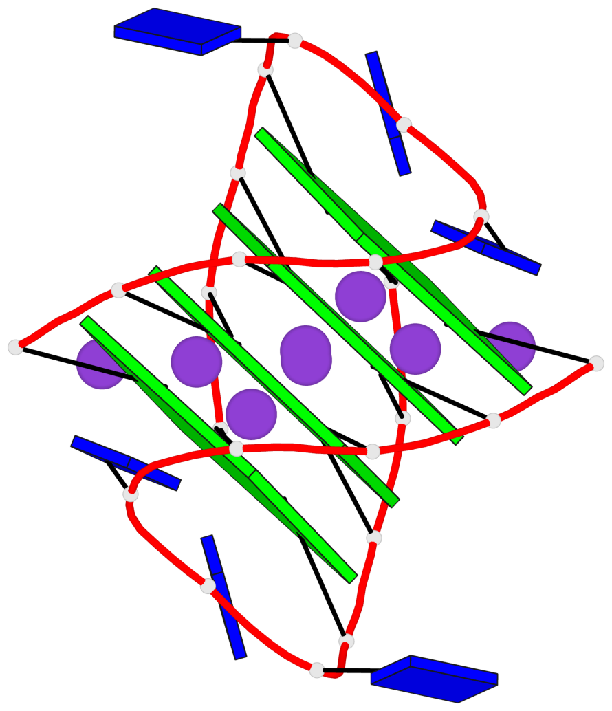
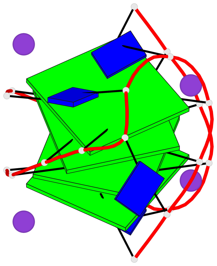
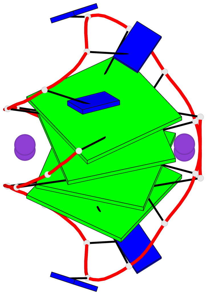

List of 4 G-tetrads
 1 glyco-bond=s-s- sugar=---- groove=wnwn planarity=0.574 type=bowl   nts=4 GGGG 1:A.DG1,1:A.DG11,2:A.DG8,2:A.DG4
 2 glyco-bond=-s-s sugar=---- groove=wnwn planarity=0.435 type=other  nts=4 GGGG 1:A.DG2,1:A.DG10,2:A.DG9,2:A.DG3
 3 glyco-bond=s-s- sugar=---- groove=wnwn planarity=0.435 type=other  nts=4 GGGG 1:A.DG3,1:A.DG9,2:A.DG10,2:A.DG2
 4 glyco-bond=-s-s sugar=---- groove=wnwn planarity=0.574 type=bowl   nts=4 GGGG 1:A.DG4,1:A.DG8,2:A.DG11,2:A.DG1
List of 1 G4-helix
In DSSR, a G4-helix is defined by stacking interactions of G-tetrads, regardless of backbone connectivity, and may contain more than one G4-stem.
Helix#1, 4 G-tetrad layers, inter-molecular, with 1 stem
 |
1 glyco-bond=s-s- sugar=---- groove=wnwn Major-->WC nts=4 GGGG 1:A.DG1,1:A.DG11,2:A.DG8,2:A.DG4 2 glyco-bond=-s-s sugar=---- groove=wnwn WC-->Major nts=4 GGGG 1:A.DG2,1:A.DG10,2:A.DG9,2:A.DG3 3 glyco-bond=s-s- sugar=---- groove=wnwn Major-->WC nts=4 GGGG 1:A.DG3,1:A.DG9,2:A.DG10,2:A.DG2 4 glyco-bond=-s-s sugar=---- groove=wnwn WC-->Major nts=4 GGGG 1:A.DG4,1:A.DG8,2:A.DG11,2:A.DG1 step#1 mm(<>,outward) area=14.99 rise=3.58 twist=16.1 step#2 pp(><,inward) area=19.33 rise=3.84 twist=33.3 step#3 mm(<>,outward) area=14.99 rise=3.58 twist=16.1 strand#1 DNA glyco-bond=s-s- sugar=---- nts=4 GGGG 1:A.DG1,1:A.DG2,1:A.DG3,1:A.DG4 strand#2 DNA glyco-bond=-s-s sugar=---- nts=4 GGGG 1:A.DG11,1:A.DG10,1:A.DG9,1:A.DG8 strand#3 DNA glyco-bond=s-s- sugar=---- nts=4 GGGG 2:A.DG8,2:A.DG9,2:A.DG10,2:A.DG11 strand#4 DNA glyco-bond=-s-s sugar=---- nts=4 GGGG 2:A.DG4,2:A.DG3,2:A.DG2,2:A.DG1 |
| 3 stacking diagrams | |
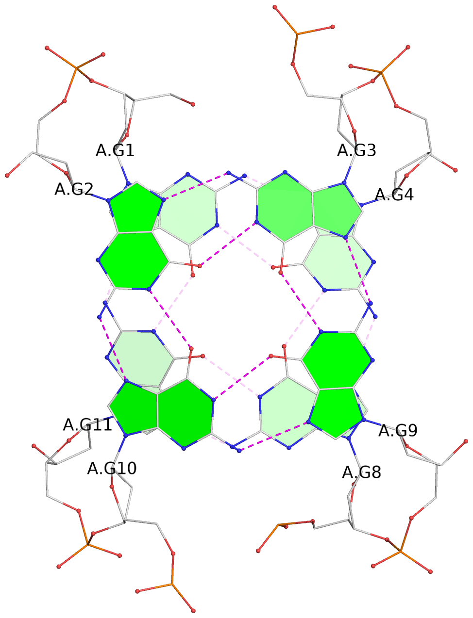 |
1 glyco-bond=s-s- sugar=---- groove=wnwn Major-->WC nts=4 GGGG 1:A.DG1,1:A.DG11,2:A.DG8,2:A.DG4 |
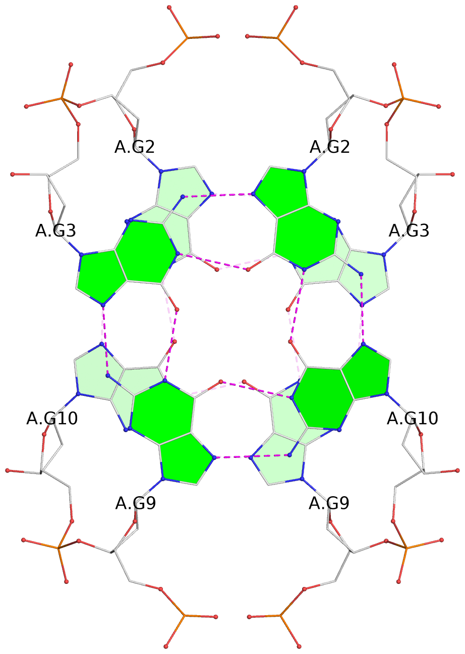 |
2 glyco-bond=-s-s sugar=---- groove=wnwn WC-->Major nts=4 GGGG 1:A.DG2,1:A.DG10,2:A.DG9,2:A.DG3 |
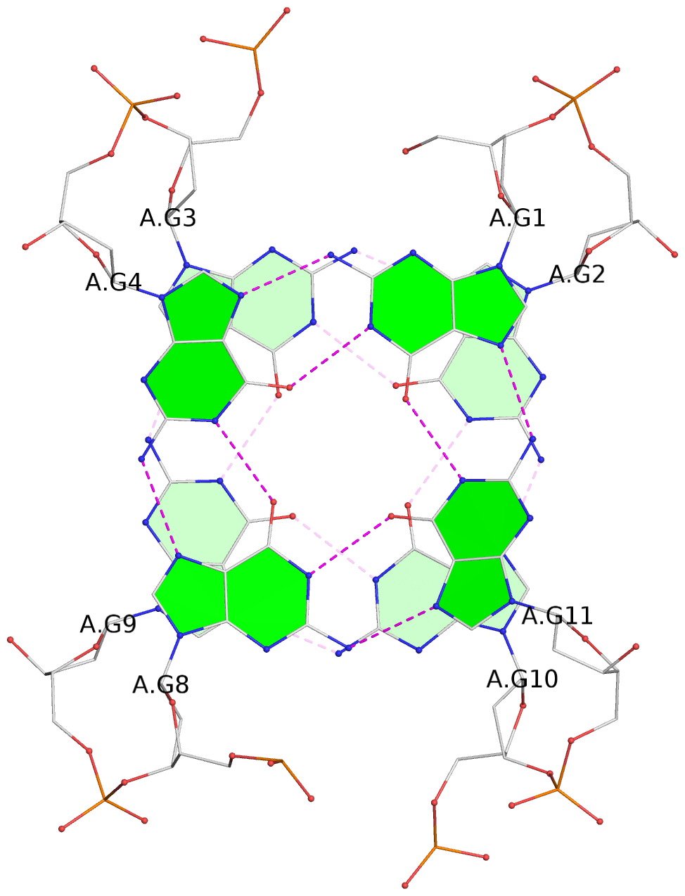 |
3 glyco-bond=s-s- sugar=---- groove=wnwn Major-->WC nts=4 GGGG 1:A.DG3,1:A.DG9,2:A.DG10,2:A.DG2 |
List of 1 G4-stem
In DSSR, a G4-stem is defined as a G4-helix with backbone connectivity. Bulges are also allowed along each of the four strands.
Stem#1, 4 G-tetrad layers, 2 loops, inter-molecular, UDUD, anti-parallel, (2+2)
 |
1 glyco-bond=s-s- sugar=---- groove=wnwn Major-->WC nts=4 GGGG 1:A.DG1,1:A.DG11,2:A.DG8,2:A.DG4 2 glyco-bond=-s-s sugar=---- groove=wnwn WC-->Major nts=4 GGGG 1:A.DG2,1:A.DG10,2:A.DG9,2:A.DG3 3 glyco-bond=s-s- sugar=---- groove=wnwn Major-->WC nts=4 GGGG 1:A.DG3,1:A.DG9,2:A.DG10,2:A.DG2 4 glyco-bond=-s-s sugar=---- groove=wnwn WC-->Major nts=4 GGGG 1:A.DG4,1:A.DG8,2:A.DG11,2:A.DG1 step#1 mm(<>,outward) area=14.99 rise=3.58 twist=16.1 step#2 pp(><,inward) area=19.33 rise=3.84 twist=33.3 step#3 mm(<>,outward) area=14.99 rise=3.58 twist=16.1 strand#1 U DNA glyco-bond=s-s- sugar=---- nts=4 GGGG 1:A.DG1,1:A.DG2,1:A.DG3,1:A.DG4 strand#2 D DNA glyco-bond=-s-s sugar=---- nts=4 GGGG 1:A.DG11,1:A.DG10,1:A.DG9,1:A.DG8 strand#3 U DNA glyco-bond=s-s- sugar=---- nts=4 GGGG 2:A.DG8,2:A.DG9,2:A.DG10,2:A.DG11 strand#4 D DNA glyco-bond=-s-s sugar=---- nts=4 GGGG 2:A.DG4,2:A.DG3,2:A.DG2,2:A.DG1 loop#1 type=lateral strands=[#1,#2] nts=3 TTT 1:A.DT5,1:A.DT6,1:A.DT7 loop#2 type=lateral strands=[#4,#3] nts=3 TTT 2:A.DT5,2:A.DT6,2:A.DT7 |