Detailed DSSR results for the G-quadruplex: PDB entry 2jpz
Created and maintained by Xiang-Jun Lu <xiangjun@x3dna.org>
Citation: Please cite the NAR'20 DSSR-PyMOL schematics paper and/or the NAR'15 DSSR method paper.
Summary information
- PDB id
- 2jpz
- Class
- DNA
- Method
- NMR
- Summary
- Human telomere DNA quadruplex structure in k+ solution hybrid-2 form
- Reference
- Dai J, Carver M, Punchihewa C, Jones RA, Yang D (2007): "Structure of the Hybrid-2 type intramolecular human telomeric G-quadruplex in K+ solution: insights into structure polymorphism of the human telomeric sequence." Nucleic Acids Res., 35, 4927-4940. doi: 10.1093/nar/gkm522.
- Abstract
- Formation of the G-quadruplex in the human telomeric sequence can inhibit the activity of telomerase, thus the intramolecular telomeric G-quadruplexes have been considered as an attractive anticancer target. Information of intramolecular telomeric G-quadruplex structures formed under physiological conditions is important for structure-based drug design. Here, we report the first structure of the major intramolecular G-quadruplex formed in a native, non-modified human telomeric sequence in K(+) solution. This is a hybrid-type mixed parallel/antiparallel-G-stranded G-quadruplex, one end of which is covered by a novel T:A:T triple capping structure. This structure (Hybrid-2) and the previously reported Hybrid-1 structure differ in their loop arrangements, strand orientations and capping structures. The distinct capping structures appear to be crucial for the favored formation of the specific hybrid-type intramolecular telomeric G-quadruplexes, and may provide specific binding sites for drug targeting. Our study also shows that while the hybrid-type G-quadruplexes appear to be the major conformations in K(+) solution, human telomeric sequences are always in equilibrium between Hybrid-1 and Hybrid-2 structures, which is largely determined by the 3'-flanking sequence. Furthermore, both hybrid-type G-quadruplexes suggest a straightforward means for multimer formation with effective packing in the human telomeric sequence and provide important implications for drug targeting of G-quadruplexes in human telomeres.
- G4 notes
- 3 G-tetrads, 1 G4 helix, 1 G4 stem, 3(-Lw-Ln-P), hybrid-2(3+1), UDUU
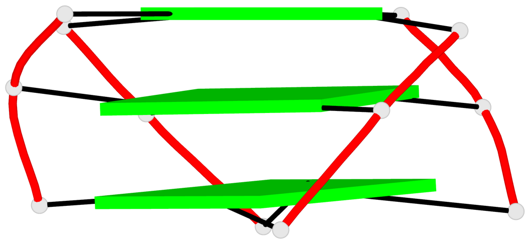
Base-block schematics in six views
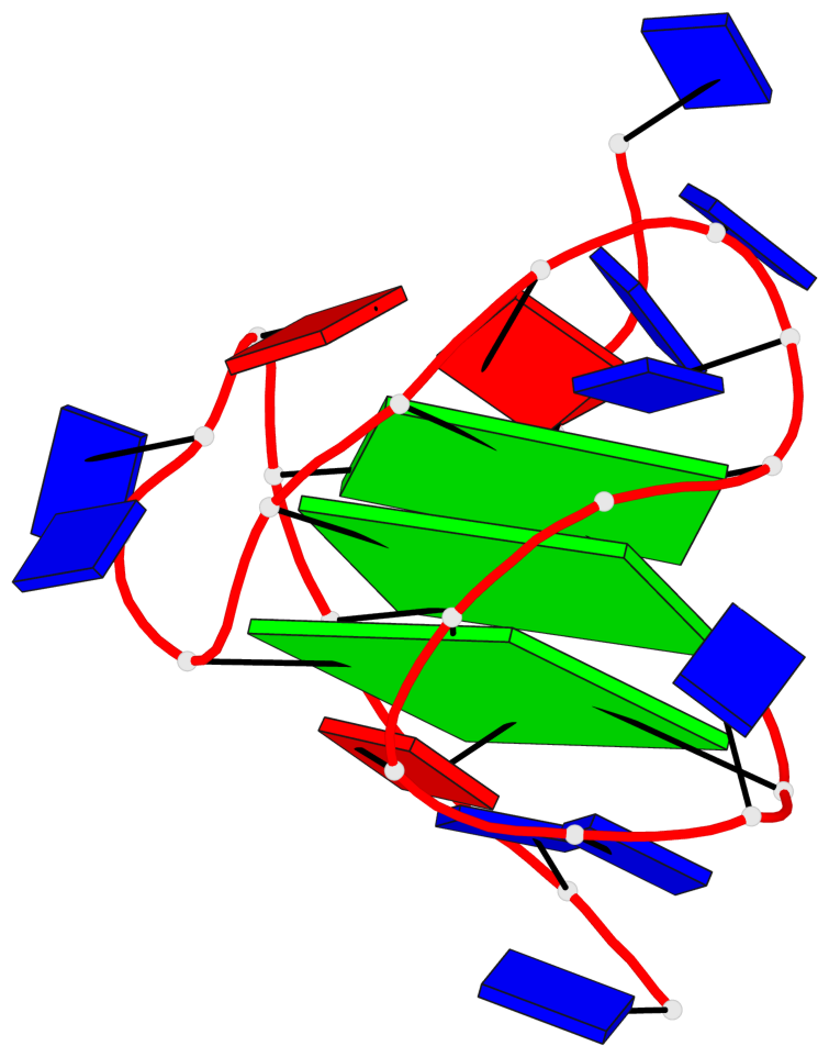
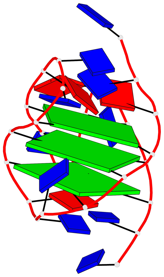
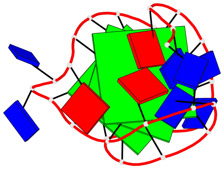
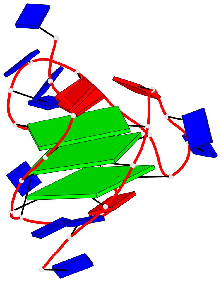
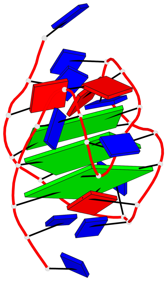
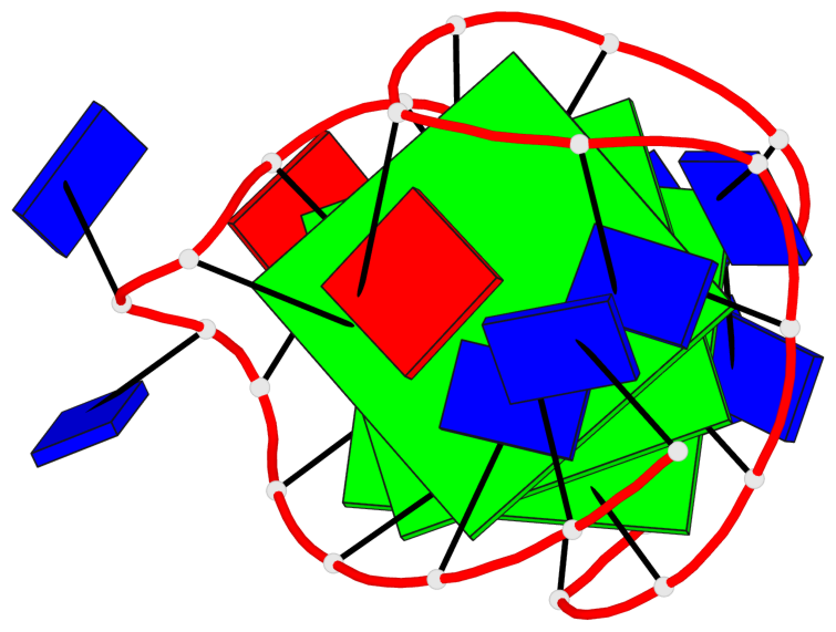
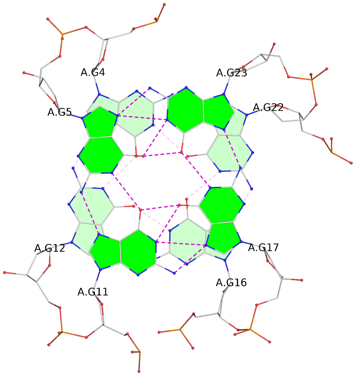
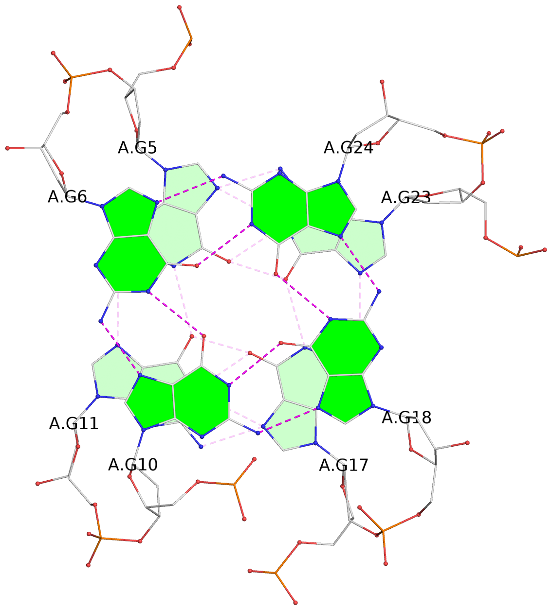
List of 3 G-tetrads
 1 glyco-bond=s-ss sugar=-.-- groove=wn-- planarity=0.189 type=other  nts=4 GGGG A.DG4,A.DG12,A.DG16,A.DG22
 2 glyco-bond=-s-- sugar=.-.. groove=wn-- planarity=0.079 type=planar nts=4 GGGG A.DG5,A.DG11,A.DG17,A.DG23
 3 glyco-bond=-s-- sugar=.-.. groove=wn-- planarity=0.085 type=planar nts=4 GGGG A.DG6,A.DG10,A.DG18,A.DG24
List of 1 G4-helix
In DSSR, a G4-helix is defined by stacking interactions of G-tetrads, regardless of backbone connectivity, and may contain more than one G4-stem.
Helix#1, 3 G-tetrad layers, INTRA-molecular, with 1 stem
 |
1 glyco-bond=s-ss sugar=-.-- groove=wn-- Major-->WC nts=4 GGGG A.DG4,A.DG12,A.DG16,A.DG22 2 glyco-bond=-s-- sugar=.-.. groove=wn-- WC-->Major nts=4 GGGG A.DG5,A.DG11,A.DG17,A.DG23 3 glyco-bond=-s-- sugar=.-.. groove=wn-- WC-->Major nts=4 GGGG A.DG6,A.DG10,A.DG18,A.DG24 step#1 mm(<>,outward) area=10.22 rise=3.44 twist=24.0 step#2 pm(>>,forward) area=16.90 rise=3.72 twist=22.7 strand#1 DNA glyco-bond=s-- sugar=-.. nts=3 GGG A.DG4,A.DG5,A.DG6 strand#2 DNA glyco-bond=-ss sugar=.-- nts=3 GGG A.DG12,A.DG11,A.DG10 strand#3 DNA glyco-bond=s-- sugar=-.. nts=3 GGG A.DG16,A.DG17,A.DG18 strand#4 DNA glyco-bond=s-- sugar=-.. nts=3 GGG A.DG22,A.DG23,A.DG24 |
| 2 stacking diagrams | |
 |
1 glyco-bond=s-ss sugar=-.-- groove=wn-- Major-->WC nts=4 GGGG A.DG4,A.DG12,A.DG16,A.DG22 |
 |
2 glyco-bond=-s-- sugar=.-.. groove=wn-- WC-->Major nts=4 GGGG A.DG5,A.DG11,A.DG17,A.DG23 |
List of 1 G4-stem
In DSSR, a G4-stem is defined as a G4-helix with backbone connectivity. Bulges are also allowed along each of the four strands.
Stem#1, 3 G-tetrad layers, 3 loops, INTRA-molecular, UDUU, hybrid-(mixed), 3(-Lw-Ln-P), hybrid-2(3+1)
 |
1 glyco-bond=s-ss sugar=-.-- groove=wn-- Major-->WC nts=4 GGGG A.DG4,A.DG12,A.DG16,A.DG22 2 glyco-bond=-s-- sugar=.-.. groove=wn-- WC-->Major nts=4 GGGG A.DG5,A.DG11,A.DG17,A.DG23 3 glyco-bond=-s-- sugar=.-.. groove=wn-- WC-->Major nts=4 GGGG A.DG6,A.DG10,A.DG18,A.DG24 step#1 mm(<>,outward) area=10.22 rise=3.44 twist=24.0 step#2 pm(>>,forward) area=16.90 rise=3.72 twist=22.7 strand#1 U DNA glyco-bond=s-- sugar=-.. nts=3 GGG A.DG4,A.DG5,A.DG6 strand#2 D DNA glyco-bond=-ss sugar=.-- nts=3 GGG A.DG12,A.DG11,A.DG10 strand#3 U DNA glyco-bond=s-- sugar=-.. nts=3 GGG A.DG16,A.DG17,A.DG18 strand#4 U DNA glyco-bond=s-- sugar=-.. nts=3 GGG A.DG22,A.DG23,A.DG24 loop#1 type=lateral strands=[#1,#2] nts=3 TTA A.DT7,A.DT8,A.DA9 loop#2 type=lateral strands=[#2,#3] nts=3 TTA A.DT13,A.DT14,A.DA15 loop#3 type=propeller strands=[#3,#4] nts=3 TTA A.DT19,A.DT20,A.DA21 |