Detailed DSSR results for the G-quadruplex: PDB entry 3qlp
Created and maintained by Xiang-Jun Lu <xiangjun@x3dna.org>
Citation: Please cite the NAR'20 DSSR-PyMOL schematics paper and/or the NAR'15 DSSR method paper.
Summary information
- PDB id
- 3qlp
- Class
- hydrolase-hydrolase inhibitor-DNA
- Method
- X-ray (2.14 Å)
- Summary
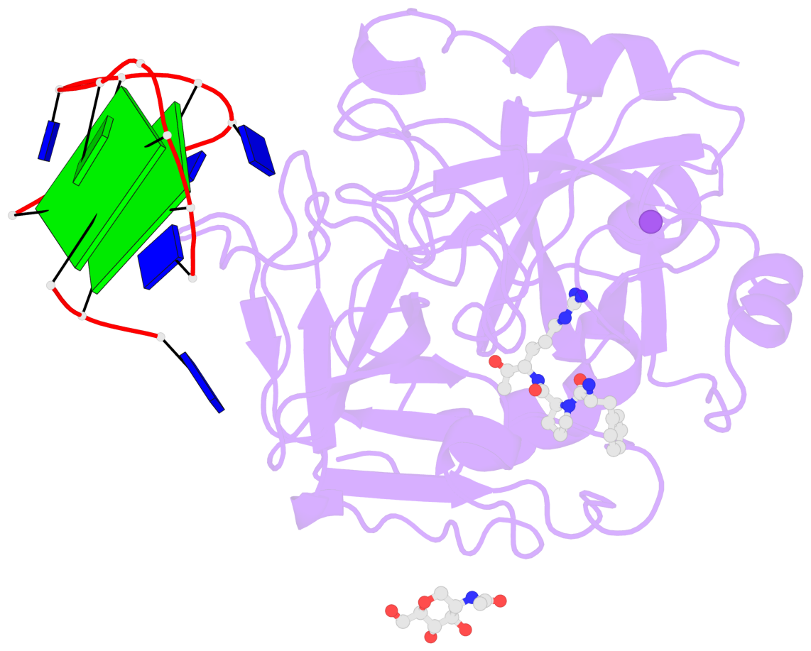
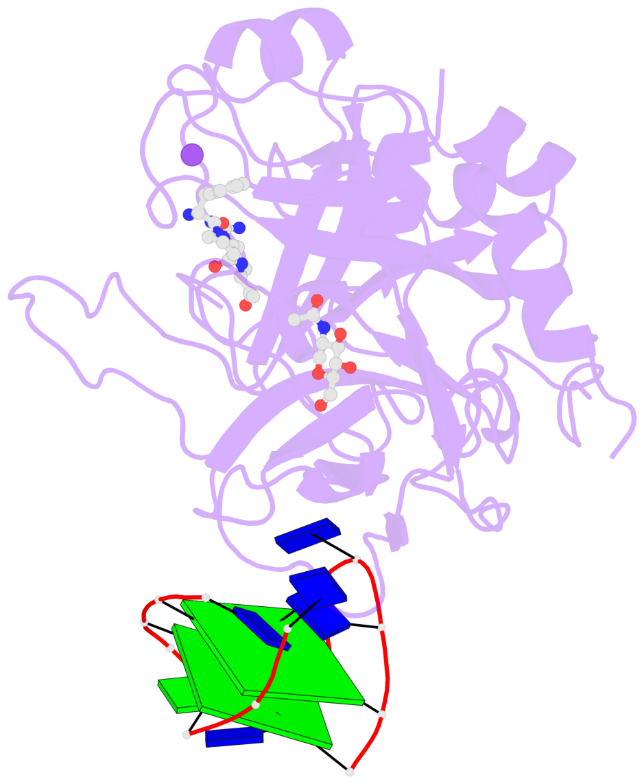
- X-ray structure of the complex between human alpha thrombin and a modified thrombin binding aptamer (mtba)
- Reference
- Russo Krauss I, Merlino A, Giancola C, Randazzo A, Mazzarella L, Sica F (2011): "Thrombin-aptamer recognition: a revealed ambiguity." Nucleic Acids Res., 39, 7858-7867. doi: 10.1093/nar/gkr522.
- Abstract
- Aptamers are structured oligonucleotides that recognize molecular targets and can function as direct protein inhibitors. The best-known example is the thrombin-binding aptamer, TBA, a single-stranded 15-mer DNA that inhibits the activity of thrombin, the key enzyme of coagulation cascade. TBA folds as a G-quadruplex structure, as proved by its NMR structure. The X-ray structure of the complex between TBA and human α-thrombin was solved at 2.9-Å resolution, but did not provide details of the aptamer conformation and the interactions with the protein molecule. TBA is rapidly processed by nucleases. To improve the properties of TBA, a number of modified analogs have been produced. In particular, a modified TBA containing a 5'-5' polarity inversion site, mTBA, has higher stability and higher affinity toward thrombin with respect to TBA, although it has a lower inhibitory activity. We present the crystal structure of the thrombin-mTBA complex at 2.15-Å resolution; the resulting model eventually provides a clear picture of thrombin-aptamers interaction, and also highlights the structural bases of the different properties of TBA and mTBA. Our findings open the way for a rational design of modified aptamers with improved potency as anticoagulant drugs.
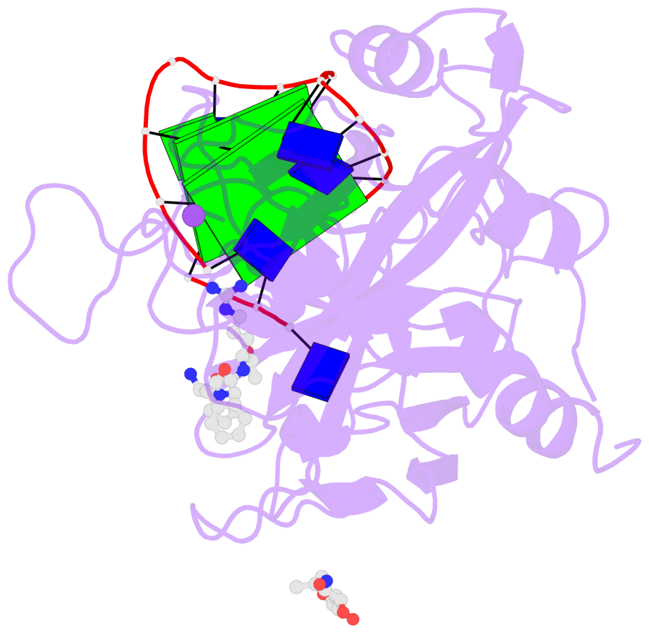
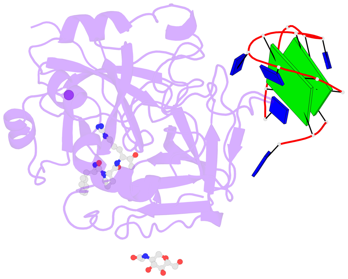
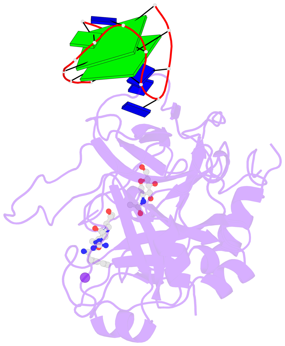
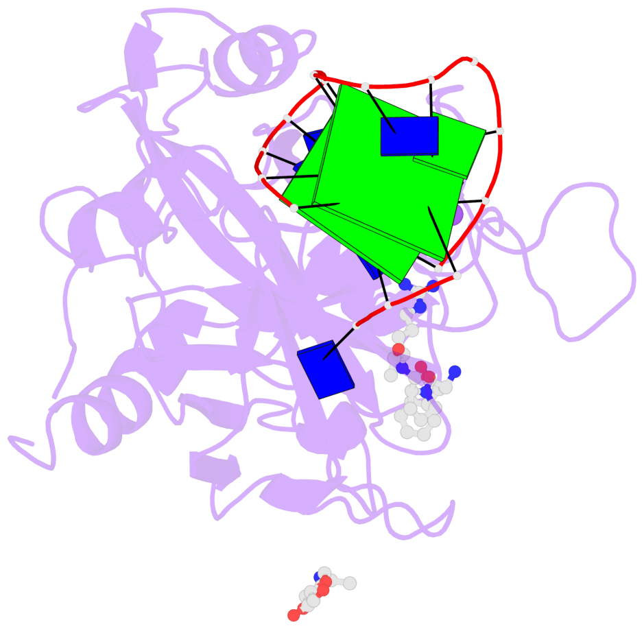
- G4 notes
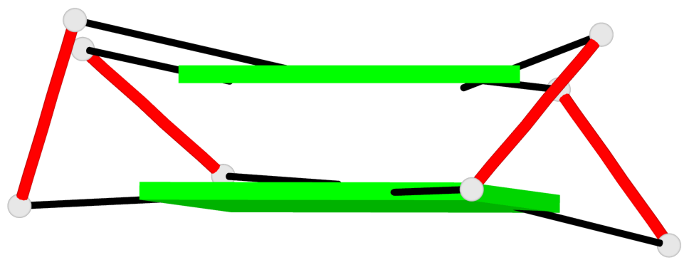
- 2 G-tetrads, 1 G4 helix, 1 G4 stem, 2(+Lm+Lw+Ln), UD3(1+3), DDUD
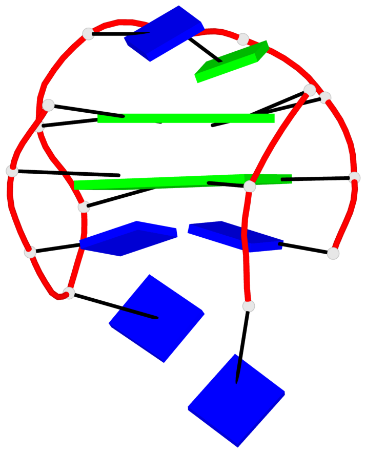
Base-block schematics in six views
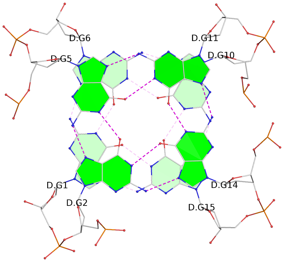
List of 2 G-tetrads
 1 glyco-bond=--s- sugar=---- groove=-wn- planarity=0.267 type=other  nts=4 GGGG D.DG1,D.DG6,D.DG10,D.DG15
 2 glyco-bond=ss-s sugar=---- groove=-wn- planarity=0.236 type=other  nts=4 GGGG D.DG2,D.DG5,D.DG11,D.DG14
List of 1 G4-helix
In DSSR, a G4-helix is defined by stacking interactions of G-tetrads, regardless of backbone connectivity, and may contain more than one G4-stem.
Helix#1, 2 G-tetrad layers, INTRA-molecular, with 1 stem
 |
1 glyco-bond=--s- sugar=---- groove=-wn- WC-->Major nts=4 GGGG D.DG1,D.DG6,D.DG10,D.DG15 2 glyco-bond=ss-s sugar=---- groove=-wn- Major-->WC nts=4 GGGG D.DG2,D.DG5,D.DG11,D.DG14 step#1 mm(<>,outward) area=16.30 rise=3.45 twist=15.6 strand#1 DNA glyco-bond=-s sugar=-- nts=2 GG D.DG1,D.DG2 strand#2 DNA glyco-bond=-s sugar=-- nts=2 GG D.DG6,D.DG5 strand#3 DNA glyco-bond=s- sugar=-- nts=2 GG D.DG10,D.DG11 strand#4 DNA glyco-bond=-s sugar=-- nts=2 GG D.DG15,D.DG14 |
| 1 stacking diagram | |
 |
1 glyco-bond=--s- sugar=---- groove=-wn- WC-->Major nts=4 GGGG D.DG1,D.DG6,D.DG10,D.DG15 |
List of 1 G4-stem
In DSSR, a G4-stem is defined as a G4-helix with backbone connectivity. Bulges are also allowed along each of the four strands.
Stem#1, 2 G-tetrad layers, 3 loops, INTRA-molecular, DDUD, hybrid-(mixed), 2(+Lm+Lw+Ln), UD3(1+3)
 |
1 glyco-bond=--s- sugar=---- groove=-nw- Major-->WC nts=4 GGGG D.DG1,D.DG15,D.DG10,D.DG6 2 glyco-bond=ss-s sugar=---- groove=-nw- WC-->Major nts=4 GGGG D.DG2,D.DG14,D.DG11,D.DG5 step#1 mm(<>,outward) area=16.30 rise=3.45 twist=15.6 strand#1 D DNA glyco-bond=-s sugar=-- nts=2 GG D.DG1,D.DG2 strand#2 D DNA glyco-bond=-s sugar=-- nts=2 GG D.DG15,D.DG14 strand#3 U DNA glyco-bond=s- sugar=-- nts=2 GG D.DG10,D.DG11 strand#4 D DNA glyco-bond=-s sugar=-- nts=2 GG D.DG6,D.DG5 loop#1 type=lateral strands=[#1,#4] nts=2 TT D.DT3,D.DT4 loop#2 type=lateral strands=[#4,#3] nts=3 TGT D.DT7,D.DG8,D.DT9 loop#3 type=lateral strands=[#3,#2] nts=2 TT D.DT12,D.DT13 |