Detailed DSSR results for the G-quadruplex: PDB entry 4dih
Created and maintained by Xiang-Jun Lu <xiangjun@x3dna.org>
Citation: Please cite the NAR'20 DSSR-PyMOL schematics paper and/or the NAR'15 DSSR method paper.
Summary information
- PDB id
- 4dih
- Class
- hydrolase-hydrolase inhibitor-DNA
- Method
- X-ray (1.8 Å)
- Summary
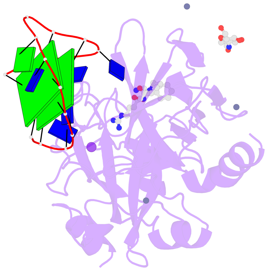
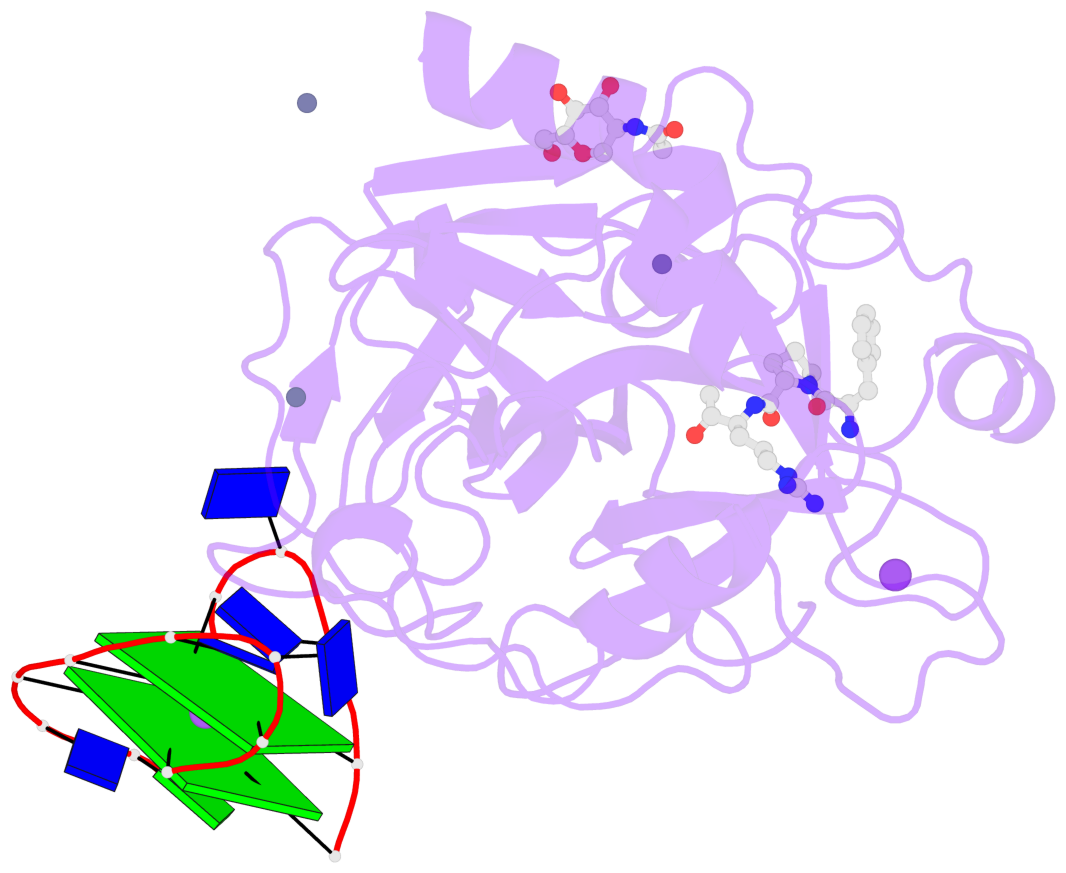
- X-ray structure of the complex between human alpha thrombin and thrombin binding aptamer in the presence of sodium ions
- Reference
- Russo Krauss I, Merlino A, Randazzo A, Novellino E, Mazzarella L, Sica F (2012): "High-resolution structures of two complexes between thrombin and thrombin-binding aptamer shed light on the role of cations in the aptamer inhibitory activity." Nucleic Acids Res., 40, 8119-8128. doi: 10.1093/nar/gks512.
- Abstract
- The G-quadruplex architecture is a peculiar structure adopted by guanine-rich oligonucleotidic sequences, and, in particular, by several aptamers, including the thrombin-binding aptamer (TBA) that has the highest inhibitory activity against human α-thrombin. A crucial role in determining structure, stability and biological properties of G-quadruplexes is played by ions. In the case of TBA, K(+) ions cause an enhancement of the aptamer clotting inhibitory activity. A detailed picture of the interactions of TBA with the protein and with the ions is still lacking, despite the importance of this aptamer in biomedical field for detection and inhibition of α-thrombin. Here, we fill this gap by presenting a high-resolution crystallographic structural characterization of the thrombin-TBA complex formed in the presence of Na(+) or K(+) and a circular dichroism study of the structural stability of the aptamer both free and complexed with α-thrombin, in the presence of the two ionic species. The results indicate that the different effects exerted by Na(+) and K(+) on the inhibitory activity of TBA are related to a subtle perturbation of a few key interactions at the protein-aptamer interface. The present data, in combination with those previously obtained on the complex between α-thrombin and a modified aptamer, may allow the design of new TBA variants with a pharmacological performance enhancement.
- G4 notes
- 2 G-tetrads, 1 G4 helix, 1 G4 stem, 2(+Ln+Lw+Ln), chair(2+2), UDUD
Base-block schematics in six views
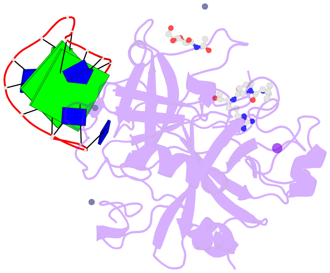
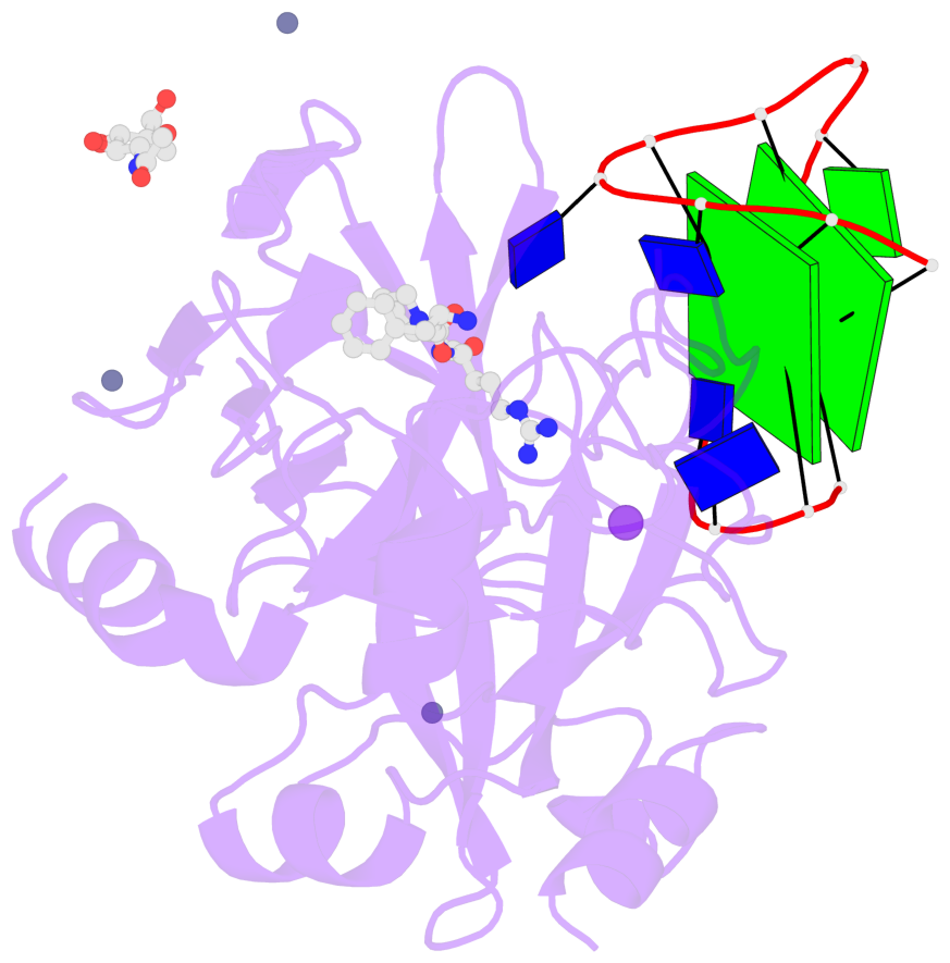
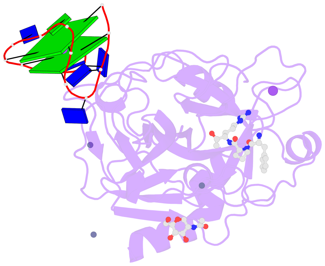
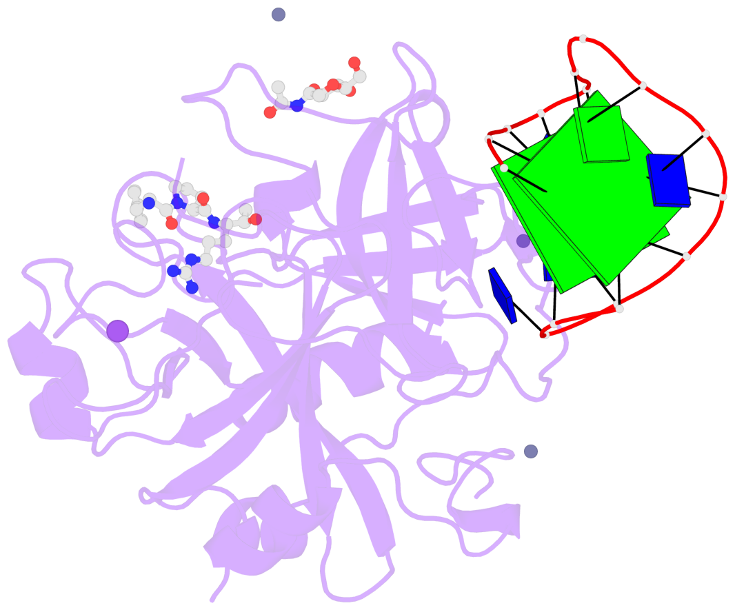
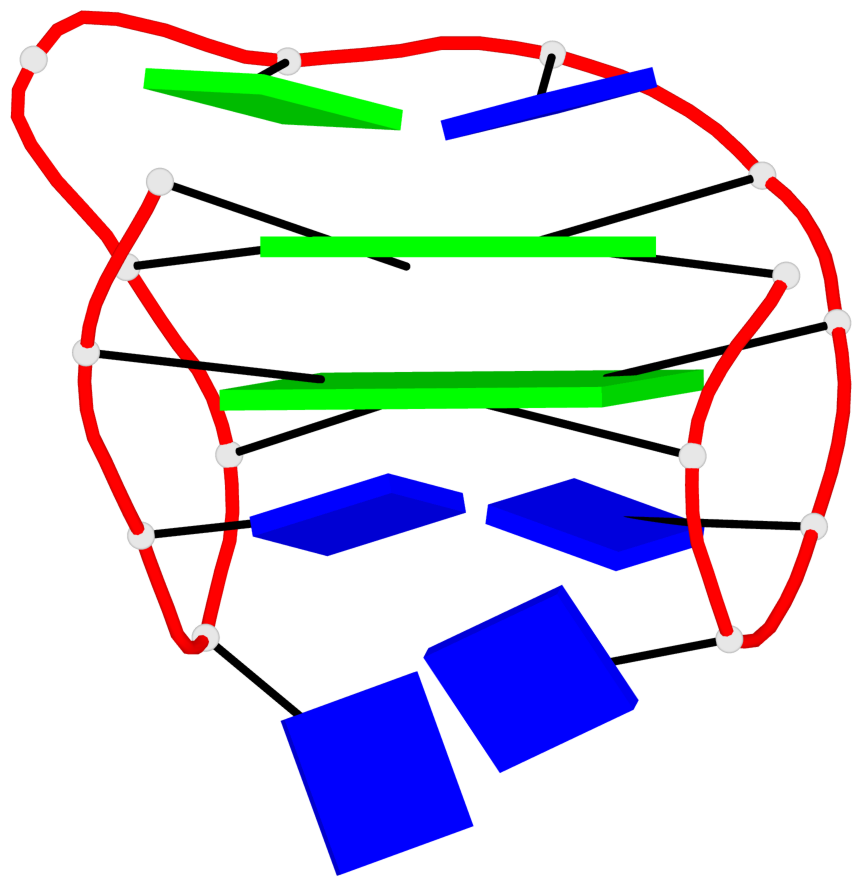
List of 2 G-tetrads
 1 glyco-bond=s-s- sugar=---- groove=wnwn planarity=0.428 type=saddle nts=4 GGGG D.DG1,D.DG15,D.DG10,D.DG6
 2 glyco-bond=-s-s sugar=---- groove=wnwn planarity=0.499 type=other  nts=4 GGGG D.DG2,D.DG14,D.DG11,D.DG5
List of 1 G4-helix
In DSSR, a G4-helix is defined by stacking interactions of G-tetrads, regardless of backbone connectivity, and may contain more than one G4-stem.
Helix#1, 2 G-tetrad layers, INTRA-molecular, with 1 stem
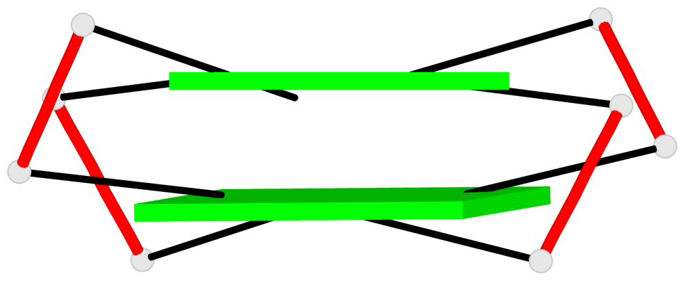 |
1 glyco-bond=s-s- sugar=---- groove=wnwn Major-->WC nts=4 GGGG D.DG1,D.DG15,D.DG10,D.DG6 2 glyco-bond=-s-s sugar=---- groove=wnwn WC-->Major nts=4 GGGG D.DG2,D.DG14,D.DG11,D.DG5 step#1 mm(<>,outward) area=15.77 rise=3.48 twist=15.0 strand#1 DNA glyco-bond=s- sugar=-- nts=2 GG D.DG1,D.DG2 strand#2 DNA glyco-bond=-s sugar=-- nts=2 GG D.DG15,D.DG14 strand#3 DNA glyco-bond=s- sugar=-- nts=2 GG D.DG10,D.DG11 strand#4 DNA glyco-bond=-s sugar=-- nts=2 GG D.DG6,D.DG5 |
| 1 stacking diagram | |
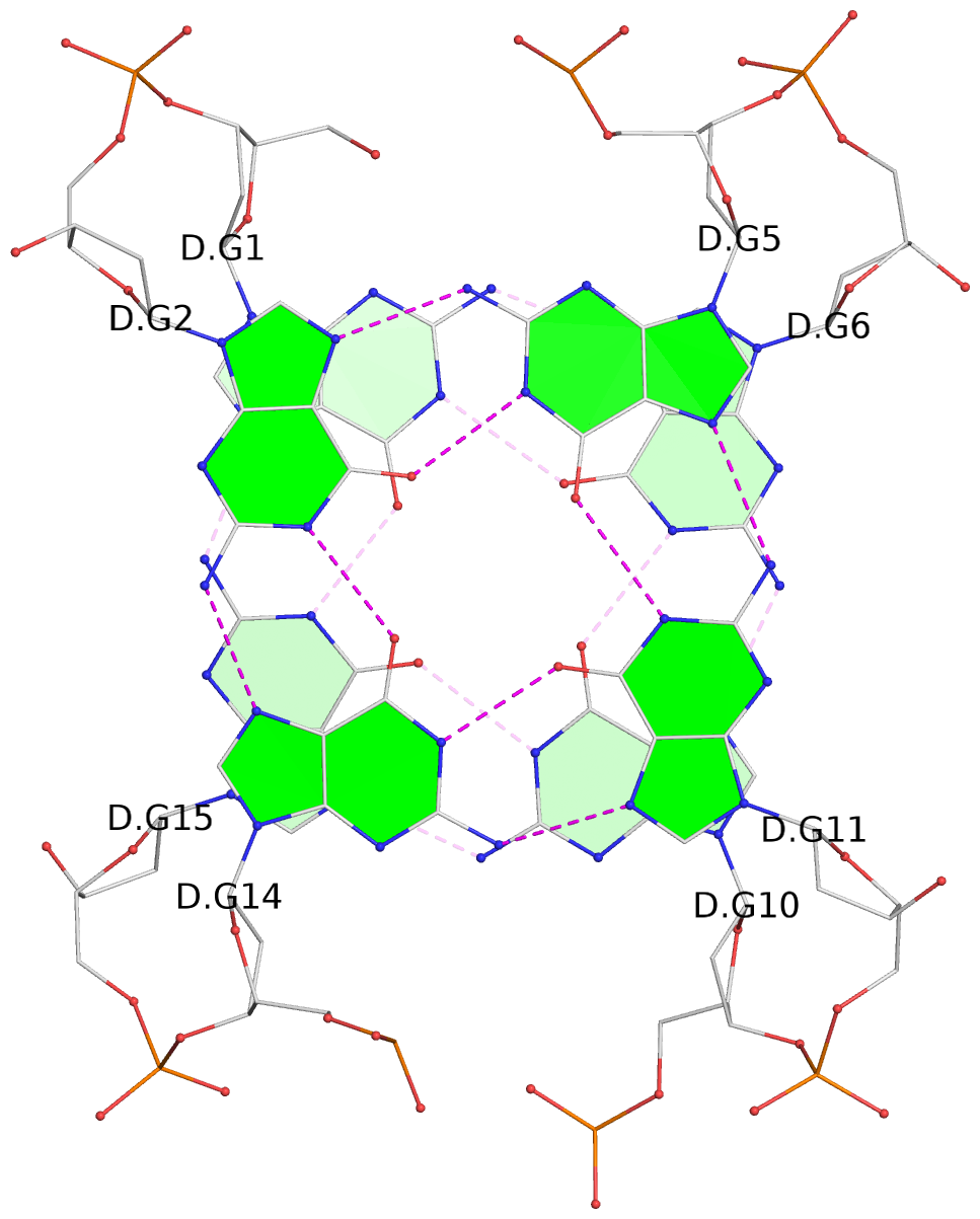 |
1 glyco-bond=s-s- sugar=---- groove=wnwn Major-->WC nts=4 GGGG D.DG1,D.DG15,D.DG10,D.DG6 |
List of 1 G4-stem
In DSSR, a G4-stem is defined as a G4-helix with backbone connectivity. Bulges are also allowed along each of the four strands.
Stem#1, 2 G-tetrad layers, 3 loops, INTRA-molecular, UDUD, anti-parallel, 2(+Ln+Lw+Ln), chair(2+2)
 |
1 glyco-bond=s-s- sugar=---- groove=wnwn Major-->WC nts=4 GGGG D.DG1,D.DG15,D.DG10,D.DG6 2 glyco-bond=-s-s sugar=---- groove=wnwn WC-->Major nts=4 GGGG D.DG2,D.DG14,D.DG11,D.DG5 step#1 mm(<>,outward) area=15.77 rise=3.48 twist=15.0 strand#1 U DNA glyco-bond=s- sugar=-- nts=2 GG D.DG1,D.DG2 strand#2 D DNA glyco-bond=-s sugar=-- nts=2 GG D.DG15,D.DG14 strand#3 U DNA glyco-bond=s- sugar=-- nts=2 GG D.DG10,D.DG11 strand#4 D DNA glyco-bond=-s sugar=-- nts=2 GG D.DG6,D.DG5 loop#1 type=lateral strands=[#1,#4] nts=2 TT D.DT3,D.DT4 loop#2 type=lateral strands=[#4,#3] nts=3 TGT D.DT7,D.DG8,D.DT9 loop#3 type=lateral strands=[#3,#2] nts=2 TT D.DT12,D.DT13 |