Detailed DSSR results for the G-quadruplex: PDB entry 5cmx
Created and maintained by Xiang-Jun Lu <xiangjun@x3dna.org>
Citation: Please cite the NAR'20 DSSR-PyMOL schematics paper and/or the NAR'15 DSSR method paper.
Summary information
- PDB id
- 5cmx
- Class
- hydrolase
- Method
- X-ray (2.98 Å)
- Summary
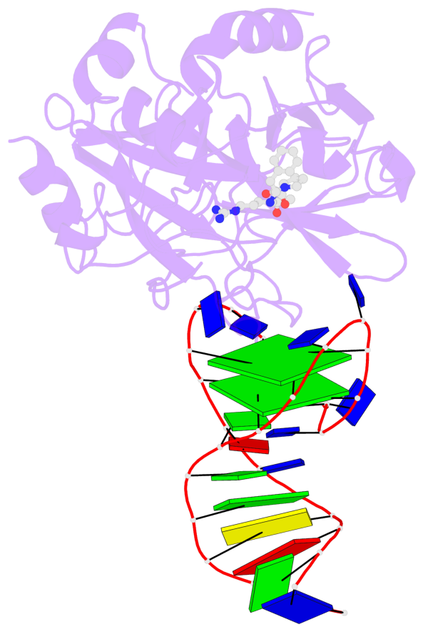
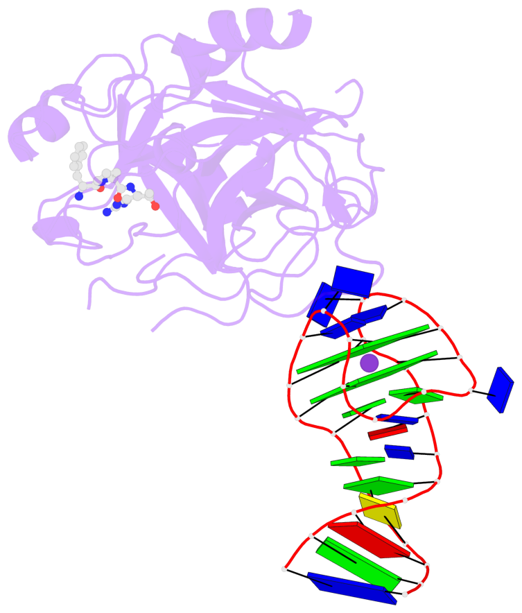
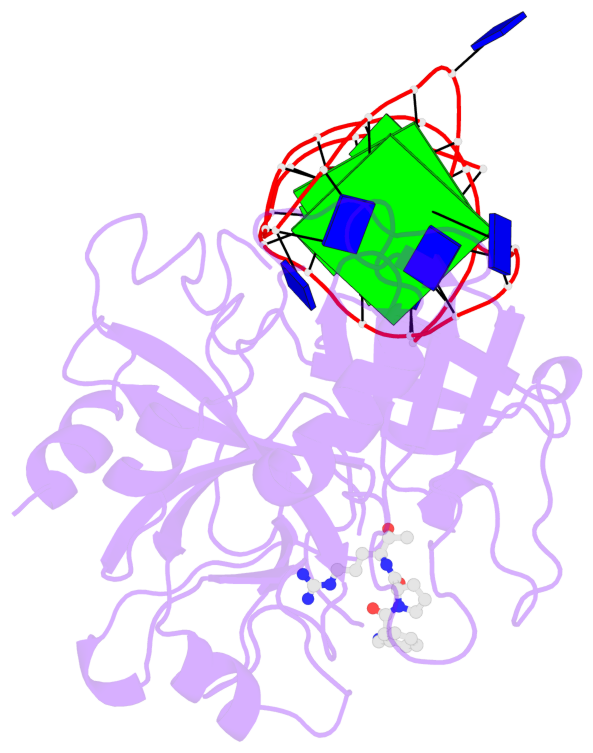
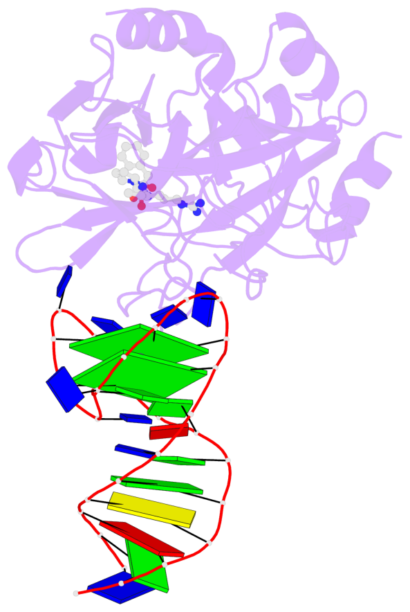
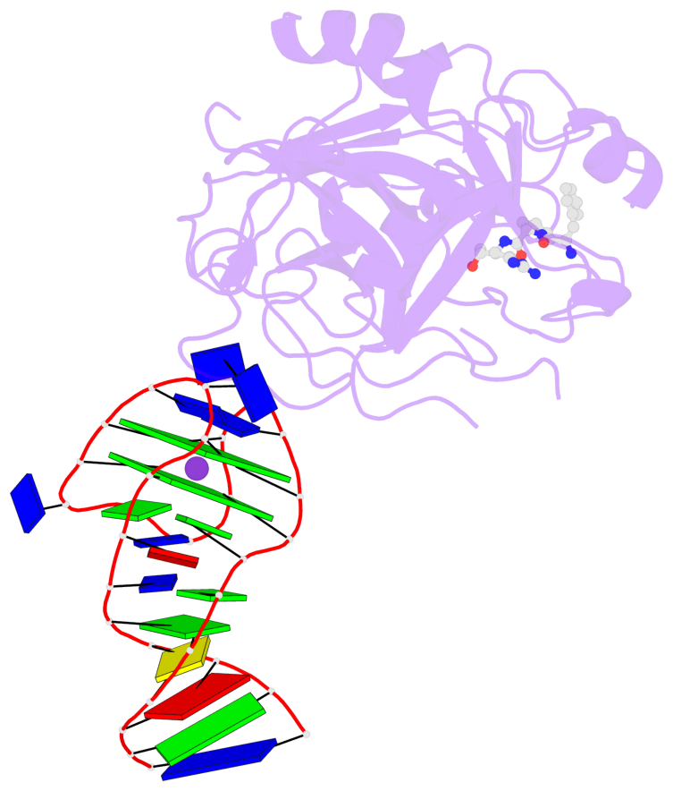
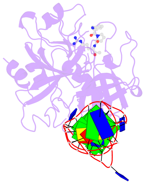
- X-ray structure of the complex between human alpha thrombin and a duplex-quadruplex 31-mer DNA aptamer
- Reference
- Russo Krauss I, Spiridonova V, Pica A, Napolitano V, Sica F (2016): "Different duplex/quadruplex junctions determine the properties of anti-thrombin aptamers with mixed folding." Nucleic Acids Res., 44, 983-991. doi: 10.1093/nar/gkv1384.
- Abstract
- Mixed duplex/quadruplex oligonucleotides have attracted great interest as therapeutic targets as well as effective biomedical aptamers. In the case of thrombin-binding aptamer (TBA), the addition of a duplex motif to the G-quadruplex module improves the aptamer resistance to biodegradation and the affinity for thrombin. In particular, the mixed oligonucleotide RE31 is significantly more effective than TBA in anticoagulation experiments and shows a slower disappearance rate in human plasma and blood. In the crystal structure of the complex with thrombin, RE31 adopts an elongated structure in which the duplex and quadruplex regions are perfectly stacked on top of each other, firmly connected by a well-structured junction. The lock-and-key shape complementarity between the TT loops of the G-quadruplex and the protein exosite I gives rise to the basic interaction that stabilizes the complex. However, our data suggest that the duplex motif may have an active role in determining the greater anti-thrombin activity in biological fluids with respect to TBA. This work gives new information on mixed oligonucleotides and highlights the importance of structural data on duplex/quadruplex junctions, which appear to be varied, unpredictable, and fundamental in determining the aptamer functional properties.
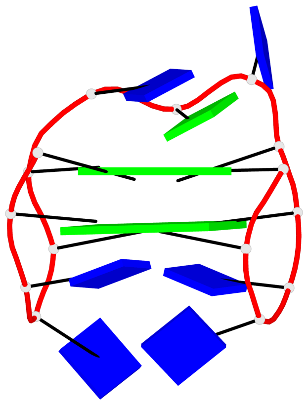
- G4 notes
- 2 G-tetrads, 1 G4 helix, 1 G4 stem, 2(+Ln+Lw+Ln), chair(2+2), UDUD
Base-block schematics in six views
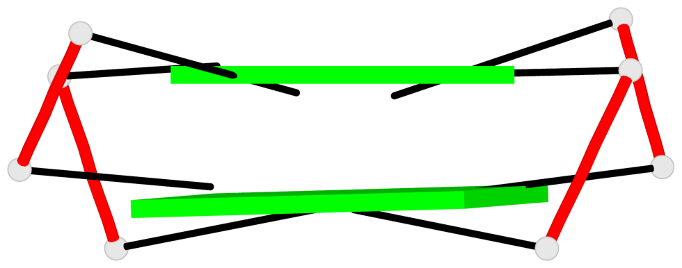
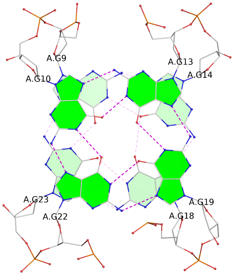
List of 2 G-tetrads
 1 glyco-bond=s-s- sugar=---- groove=wnwn planarity=0.443 type=bowl   nts=4 GGGG A.DG9,A.DG23,A.DG18,A.DG14
 2 glyco-bond=-s-s sugar=--3- groove=wnwn planarity=0.400 type=other  nts=4 GGGG A.DG10,A.DG22,A.DG19,A.DG13
List of 1 G4-helix
In DSSR, a G4-helix is defined by stacking interactions of G-tetrads, regardless of backbone connectivity, and may contain more than one G4-stem.
Helix#1, 2 G-tetrad layers, INTRA-molecular, with 1 stem
 |
1 glyco-bond=s-s- sugar=---- groove=wnwn Major-->WC nts=4 GGGG A.DG9,A.DG23,A.DG18,A.DG14 2 glyco-bond=-s-s sugar=--3- groove=wnwn WC-->Major nts=4 GGGG A.DG10,A.DG22,A.DG19,A.DG13 step#1 mm(<>,outward) area=13.73 rise=3.53 twist=14.1 strand#1 DNA glyco-bond=s- sugar=-- nts=2 GG A.DG9,A.DG10 strand#2 DNA glyco-bond=-s sugar=-- nts=2 GG A.DG23,A.DG22 strand#3 DNA glyco-bond=s- sugar=-3 nts=2 GG A.DG18,A.DG19 strand#4 DNA glyco-bond=-s sugar=-- nts=2 GG A.DG14,A.DG13 |
| 1 stacking diagram | |
 |
1 glyco-bond=s-s- sugar=---- groove=wnwn Major-->WC nts=4 GGGG A.DG9,A.DG23,A.DG18,A.DG14 |
List of 1 G4-stem
In DSSR, a G4-stem is defined as a G4-helix with backbone connectivity. Bulges are also allowed along each of the four strands.
Stem#1, 2 G-tetrad layers, 3 loops, INTRA-molecular, UDUD, anti-parallel, 2(+Ln+Lw+Ln), chair(2+2)
 |
1 glyco-bond=s-s- sugar=---- groove=wnwn Major-->WC nts=4 GGGG A.DG9,A.DG23,A.DG18,A.DG14 2 glyco-bond=-s-s sugar=--3- groove=wnwn WC-->Major nts=4 GGGG A.DG10,A.DG22,A.DG19,A.DG13 step#1 mm(<>,outward) area=13.73 rise=3.53 twist=14.1 strand#1 U DNA glyco-bond=s- sugar=-- nts=2 GG A.DG9,A.DG10 strand#2 D DNA glyco-bond=-s sugar=-- nts=2 GG A.DG23,A.DG22 strand#3 U DNA glyco-bond=s- sugar=-3 nts=2 GG A.DG18,A.DG19 strand#4 D DNA glyco-bond=-s sugar=-- nts=2 GG A.DG14,A.DG13 loop#1 type=lateral strands=[#1,#4] nts=2 TT A.DT11,A.DT12 loop#2 type=lateral strands=[#4,#3] nts=3 TGT A.DT15,A.DG16,A.DT17 loop#3 type=lateral strands=[#3,#2] nts=2 TT A.DT20,A.DT21 |