Detailed DSSR results for the G-quadruplex: PDB entry 5dwx
Created and maintained by Xiang-Jun Lu <xiangjun@x3dna.org>
Citation: Please cite the NAR'20 DSSR-PyMOL schematics paper and/or the NAR'15 DSSR method paper.
Summary information
- PDB id
- 5dwx
- Class
- DNA
- Method
- X-ray (2.71 Å)
- Summary
- Structural insights into the quadruplex-duplex 3' interface formed from a telomeric repeat - tloop
- Reference
- Russo Krauss I, Ramaswamy S, Neidle S, Haider S, Parkinson GN (2016): "Structural Insights into the Quadruplex-Duplex 3' Interface Formed from a Telomeric Repeat: A Potential Molecular Target." J.Am.Chem.Soc., 138, 1226-1233. doi: 10.1021/jacs.5b10492.
- Abstract
- We report here on an X-ray crystallographic and molecular modeling investigation into the complex 3' interface formed between putative parallel stranded G-quadruplexes and a duplex DNA sequence constructed from the human telomeric repeat sequence TTAGGG. Our crystallographic approach provides a detailed snapshot of a telomeric 3' quadruplex-duplex junction: a junction that appears to have the potential to form a unique molecular target for small molecule binding and interference with telomere-related functions. This unique target is particularly relevant as current high-affinity compounds that bind putative G-quadruplex forming sequences only rarely have a high degree of selectivity for a particular quadruplex. Here DNA junctions were assembled using different putative quadruplex-forming scaffolds linked at the 3' end to a telomeric duplex sequence and annealed to a complementary strand. We successfully generated a series of G-quadruplex-duplex containing crystals, both alone and in the presence of ligands. The structures demonstrate the formation of a parallel folded G-quadruplex and a B-form duplex DNA stacked coaxially. Most strikingly, structural data reveals the consistent formation of a TAT triad platform between the two motifs. This triad allows for a continuous stack of bases to link the quadruplex motif with the duplex region. For these crystal structures formed in the absence of ligands, the TAT triad interface occludes ligand binding at the 3' quadruplex-duplex interface, in agreement with in silico docking predictions. However, with the rearrangement of a single nucleotide, a stable pocket can be produced, thus providing an opportunity for the binding of selective molecules at the interface.
- G4 notes
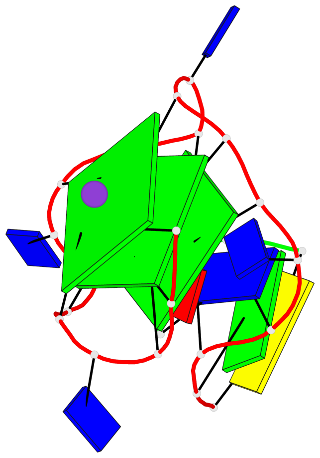
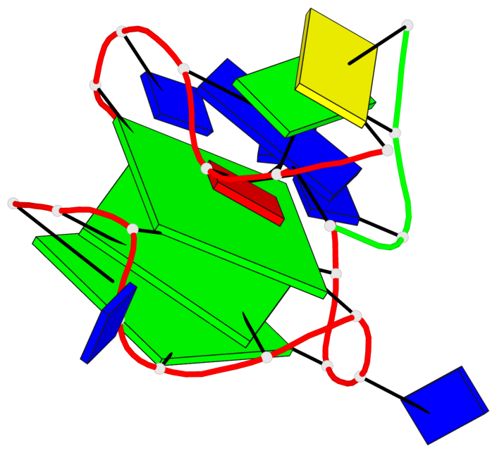
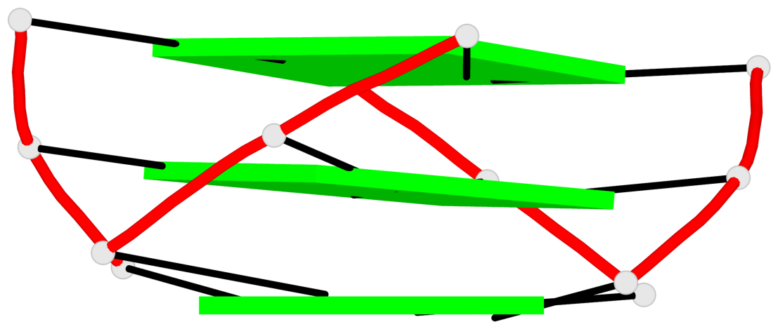
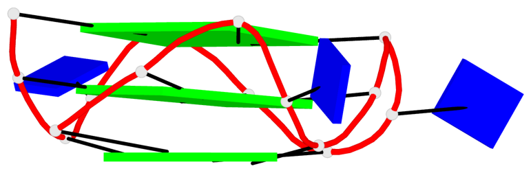
- 3 G-tetrads, 1 G4 helix, 1 G4 stem, 3(-P-P-P), parallel(4+0), UUUU
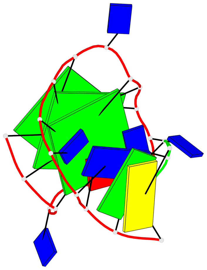
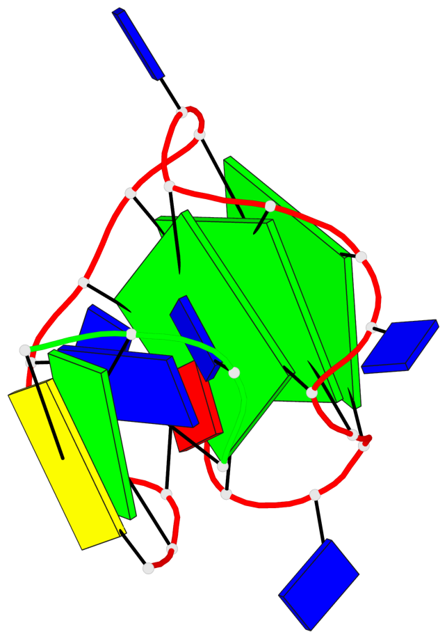
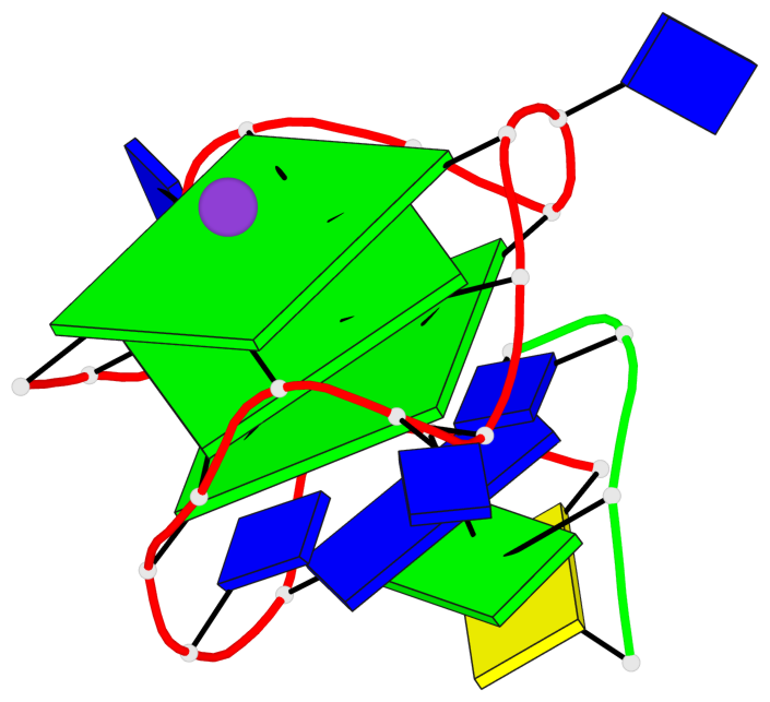
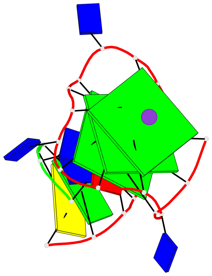
Base-block schematics in six views
List of 3 G-tetrads
 1 glyco-bond=---- sugar=---- groove=---- planarity=0.168 type=other  nts=4 GGGG A.DG1,A.DG5,A.DG9,A.DG13
 2 glyco-bond=---- sugar=---- groove=---- planarity=0.181 type=other  nts=4 GGGG A.DG2,A.DG6,A.DG10,A.DG14
 3 glyco-bond=---- sugar=---- groove=---- planarity=0.241 type=bowl   nts=4 GGGG A.DG3,A.DG7,A.DG11,A.DG15
List of 1 G4-helix
In DSSR, a G4-helix is defined by stacking interactions of G-tetrads, regardless of backbone connectivity, and may contain more than one G4-stem.
Helix#1, 3 G-tetrad layers, INTRA-molecular, with 1 stem
 |
1 glyco-bond=---- sugar=---- groove=---- WC-->Major nts=4 GGGG A.DG1,A.DG5,A.DG9,A.DG13 2 glyco-bond=---- sugar=---- groove=---- WC-->Major nts=4 GGGG A.DG2,A.DG6,A.DG10,A.DG14 3 glyco-bond=---- sugar=---- groove=---- WC-->Major nts=4 GGGG A.DG3,A.DG7,A.DG11,A.DG15 step#1 pm(>>,forward) area=11.30 rise=3.35 twist=29.9 step#2 pm(>>,forward) area=11.49 rise=3.47 twist=29.1 strand#1 DNA glyco-bond=--- sugar=--- nts=3 GGG A.DG1,A.DG2,A.DG3 strand#2 DNA glyco-bond=--- sugar=--- nts=3 GGG A.DG5,A.DG6,A.DG7 strand#3 DNA glyco-bond=--- sugar=--- nts=3 GGG A.DG9,A.DG10,A.DG11 strand#4 DNA glyco-bond=--- sugar=--- nts=3 GGG A.DG13,A.DG14,A.DG15 |
| 2 stacking diagrams | |
 |
1 glyco-bond=---- sugar=---- groove=---- WC-->Major nts=4 GGGG A.DG1,A.DG5,A.DG9,A.DG13 |
 |
2 glyco-bond=---- sugar=---- groove=---- WC-->Major nts=4 GGGG A.DG2,A.DG6,A.DG10,A.DG14 |
List of 1 G4-stem
In DSSR, a G4-stem is defined as a G4-helix with backbone connectivity. Bulges are also allowed along each of the four strands.
Stem#1, 3 G-tetrad layers, 3 loops, INTRA-molecular, UUUU, parallel, 3(-P-P-P), parallel(4+0)
 |
1 glyco-bond=---- sugar=---- groove=---- WC-->Major nts=4 GGGG A.DG1,A.DG5,A.DG9,A.DG13 2 glyco-bond=---- sugar=---- groove=---- WC-->Major nts=4 GGGG A.DG2,A.DG6,A.DG10,A.DG14 3 glyco-bond=---- sugar=---- groove=---- WC-->Major nts=4 GGGG A.DG3,A.DG7,A.DG11,A.DG15 step#1 pm(>>,forward) area=11.30 rise=3.35 twist=29.9 step#2 pm(>>,forward) area=11.49 rise=3.47 twist=29.1 strand#1 U DNA glyco-bond=--- sugar=--- nts=3 GGG A.DG1,A.DG2,A.DG3 strand#2 U DNA glyco-bond=--- sugar=--- nts=3 GGG A.DG5,A.DG6,A.DG7 strand#3 U DNA glyco-bond=--- sugar=--- nts=3 GGG A.DG9,A.DG10,A.DG11 strand#4 U DNA glyco-bond=--- sugar=--- nts=3 GGG A.DG13,A.DG14,A.DG15 loop#1 type=propeller strands=[#1,#2] nts=1 T A.DT4 loop#2 type=propeller strands=[#2,#3] nts=1 T A.DT8 loop#3 type=propeller strands=[#3,#4] nts=1 T A.DT12 |