Detailed DSSR results for the G-quadruplex: PDB entry 6gz6
Created and maintained by Xiang-Jun Lu <xiangjun@x3dna.org>
Citation: Please cite the NAR'20 DSSR-PyMOL schematics paper and/or the NAR'15 DSSR method paper.
Summary information
- PDB id
- 6gz6
- Class
- DNA
- Method
- X-ray (2.006 Å)
- Summary
- Structure of a left-handed g-quadruplex
- Reference
- Bakalar B, Heddi B, Schmitt E, Mechulam Y, Phan AT (2019): "A Minimal Sequence for Left-Handed G-Quadruplex Formation." Angew.Chem.Int.Ed.Engl., 58, 2331-2335. doi: 10.1002/anie.201812628.
- Abstract
- Recently, we observed the first example of a left-handed G-quadruplex structure formed by natural DNA, named Z-G4. We analysed the Z-G4 structure and inspected its primary 28-nt sequence in order to identify motifs that convey the unique left-handed twist. Using circular dichroism spectroscopy, NMR spectroscopy, and X-ray crystallography, we revealed a minimal sequence motif of 12 nt (GTGGTGGTGGTG) for formation of the left-handed DNA G-quadruplex, which is found to be highly abundant in the human genome. A systematic analysis of thymine loop mutations revealed a moderate sequence tolerance, which would further broaden the space of sequences prone to left-handed G-quadruplex formation.
- G4 notes
- 4 G-tetrads, 1 G4 helix
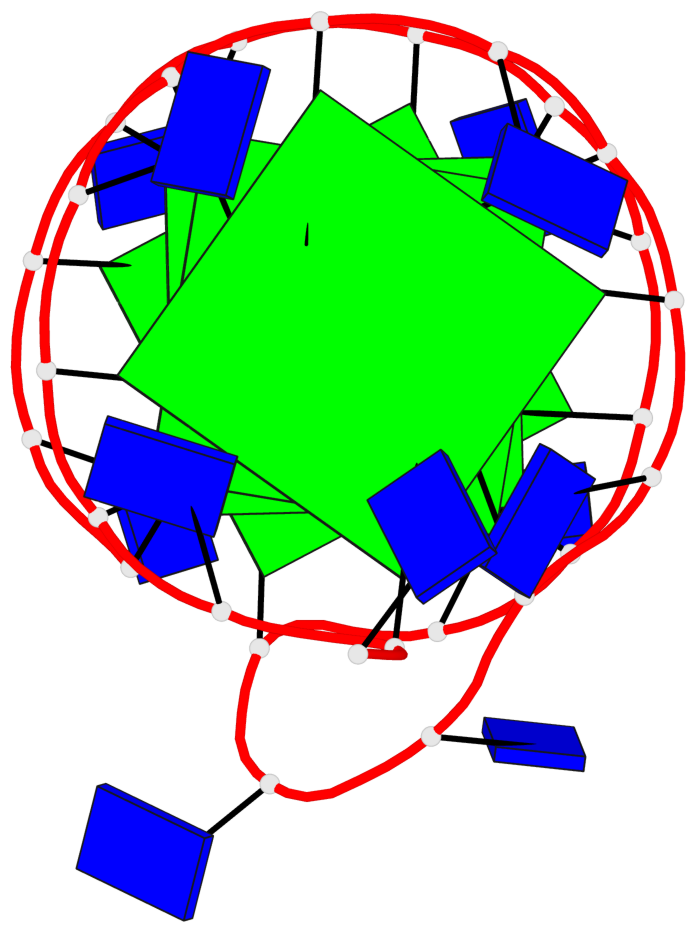
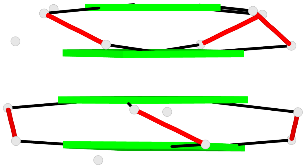
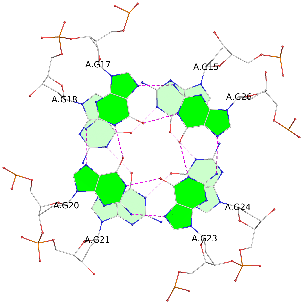
Base-block schematics in six views
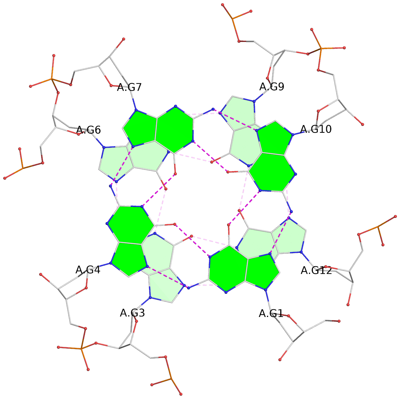
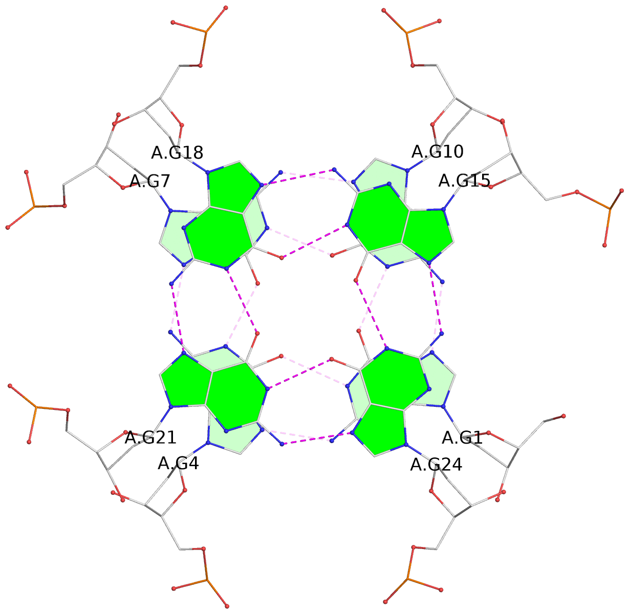
List of 4 G-tetrads
 1 glyco-bond=---- sugar=---- groove=---- planarity=0.092 type=planar nts=4 GGGG A.DG1,A.DG4,A.DG7,A.DG10
 2 glyco-bond=---- sugar=---- groove=---- planarity=0.208 type=other  nts=4 GGGG A.DG3,A.DG6,A.DG9,A.DG12
 3 glyco-bond=---- sugar=---- groove=---- planarity=0.059 type=planar nts=4 GGGG A.DG15,A.DG18,A.DG21,A.DG24
 4 glyco-bond=---- sugar=---- groove=---- planarity=0.164 type=other  nts=4 GGGG A.DG17,A.DG20,A.DG23,A.DG26
List of 1 G4-helix
In DSSR, a G4-helix is defined by stacking interactions of G-tetrads, regardless of backbone connectivity, and may contain more than one G4-stem.
Helix#1, 4 G-tetrad layers, INTRA-molecular
List of 4 non-stem G4-loops (including the two closing Gs)
 1 type=V-shaped  helix=#1 nts=4 GTGG A.DG1,A.DT2,A.DG3,A.DG4
 2* type=V-shaped  helix=#1 nts=4 GGTG A.DG9,A.DG10,A.DT11,A.DG12
 3 type=V-shaped  helix=#1 nts=4 GTGG A.DG15,A.DT16,A.DG17,A.DG18
 4* type=V-shaped  helix=#1 nts=4 GGTG A.DG23,A.DG24,A.DT25,A.DG26
