Detailed DSSR results for the G-quadruplex: PDB entry 8tns
Created and maintained by Xiang-Jun Lu <xiangjun@x3dna.org>
Citation: Please cite the NAR'20 DSSR-PyMOL schematics paper and/or the NAR'15 DSSR method paper.
Summary information
- PDB id
- 8tns
- Class
- RNA
- Method
- multiple methods: solution nmr, solution scattering
- Summary
- Solution structure of poly(ug) RNA (gu)12 g-quadruplex
- Reference
- Escobar CA, Petersen RJ, Tonelli M, Fan L, Henzler-Wildman KA, Butcher SE (2023): "Solution Structure of Poly(UG) RNA." J.Mol.Biol., 435, 168340. doi: 10.1016/j.jmb.2023.168340.
- Abstract
- Poly(UG) or "pUG" RNAs are UG or GU dinucleotide repeat sequences which are highly abundant in eukaryotes. Post-transcriptional addition of pUGs to RNA 3' ends marks mRNAs as vectors for gene silencing in C. elegans. We previously determined the crystal structure of pUG RNA bound to the ligand N-methyl mesoporphyrin IX (NMM), but the structure of free pUG RNA is unknown. Here we report the solution structure of the free pUG RNA (GU)12, as determined by nuclear magnetic resonance spectroscopy and small and wide-angle x-ray scattering (NMR-SAXS-WAXS). The low complexity sequence and 4-fold symmetry of the structure result in overlapped NMR signals that complicate chemical shift assignment. We therefore utilized single site-specific deoxyribose modifications which did not perturb the structure and introduced well-resolved methylene signals that are easily identified in NMR spectra. The solution structure ensemble has a root mean squared deviation (RMSD) of 0.62 Å and is a compact, left-handed quadruplex with a Z-form backbone, or "pUG fold." Overall, the structure agrees with the crystal structure of (GU)12 bound to NMM, indicating the pUG fold is unaltered by docking of the NMM ligand. The solution structure reveals conformational details that could not be resolved by x-ray crystallography, which explain how the pUG fold can form within longer RNAs.
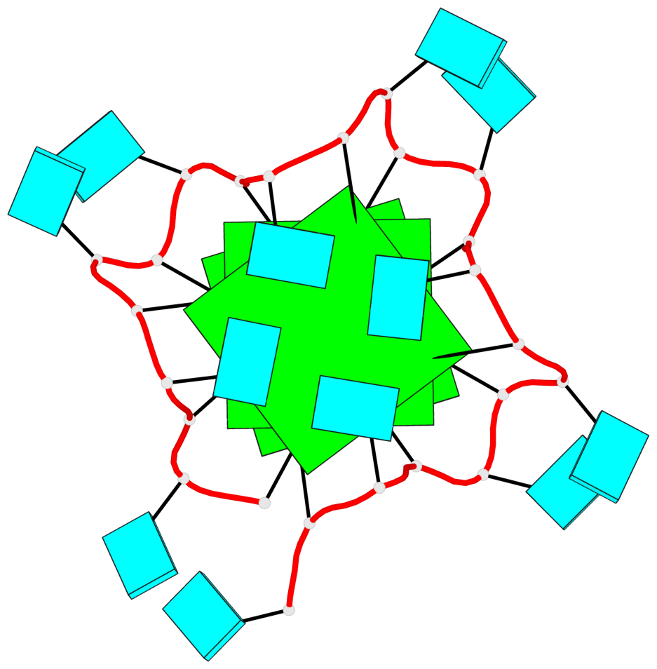
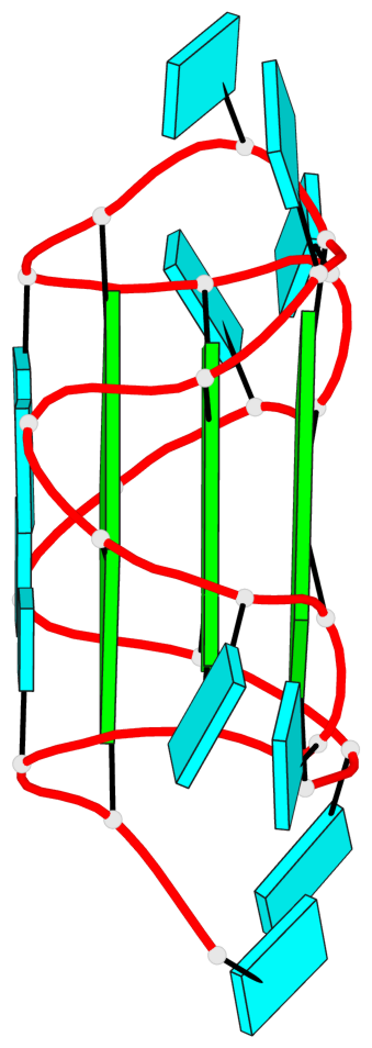
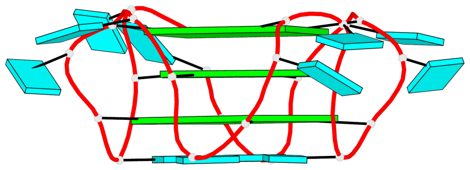
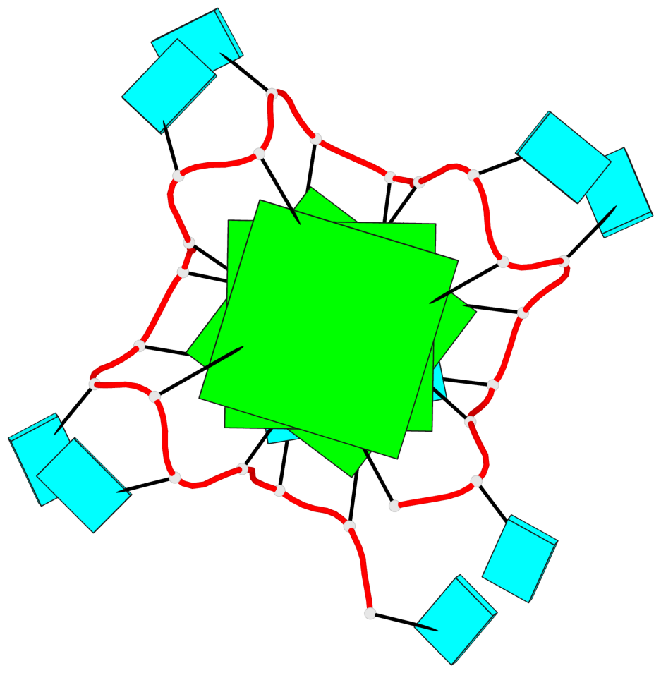
- G4 notes
- 3 G-tetrads, 1 G4 helix
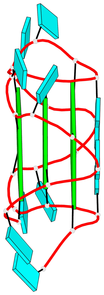
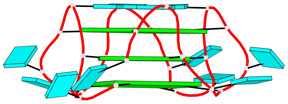
Base-block schematics in six views
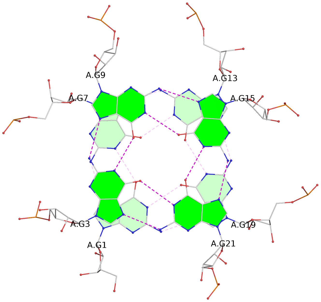
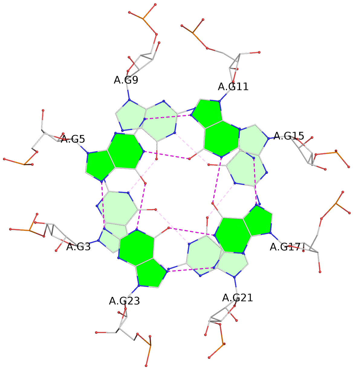
List of 3 G-tetrads
 1 glyco-bond=ssss sugar=---- groove=---- planarity=0.049 type=planar nts=4 GGGG A.G1,A.G7,A.G13,A.G19
 2 glyco-bond=ssss sugar=3333 groove=---- planarity=0.047 type=planar nts=4 GGGG A.G3,A.G21,A.G15,A.G9
 3 glyco-bond=---- sugar=---- groove=---- planarity=0.065 type=planar nts=4 GGGG A.G5,A.G11,A.G17,A.G23
List of 1 G4-helix
In DSSR, a G4-helix is defined by stacking interactions of G-tetrads, regardless of backbone connectivity, and may contain more than one G4-stem.
Helix#1, 3 G-tetrad layers, INTRA-molecular
 |
1 glyco-bond=ssss sugar=---- groove=---- Major-->WC nts=4 GGGG A.G1,A.G7,A.G13,A.G19 2 glyco-bond=ssss sugar=3333 groove=---- WC-->Major nts=4 GGGG A.G3,A.G9,A.G15,A.G21 3 glyco-bond=---- sugar=---- groove=---- WC-->Major nts=4 GGGG A.G23,A.G5,A.G11,A.G17 step#1 pp(><,inward) area=15.14 rise=3.31 twist=-16.0 step#2 mp(<<,backward) area=6.43 rise=3.60 twist=36.0 strand#1 RNA glyco-bond=ss- sugar=-3- nts=3 GGG A.G1,A.G3,A.G23 strand#2 RNA glyco-bond=ss- sugar=-3- nts=3 GGG A.G7,A.G9,A.G5 strand#3 RNA glyco-bond=ss- sugar=-3- nts=3 GGG A.G13,A.G15,A.G11 strand#4 RNA glyco-bond=ss- sugar=-3- nts=3 GGG A.G19,A.G21,A.G17 |
| 2 stacking diagrams | |
 |
1 glyco-bond=ssss sugar=---- groove=---- Major-->WC nts=4 GGGG A.G1,A.G7,A.G13,A.G19 |
 |
2 glyco-bond=ssss sugar=3333 groove=---- WC-->Major nts=4 GGGG A.G3,A.G9,A.G15,A.G21 |